Medicine - Veterinary > Veterinary colleges and laboratories > Indian journal of veterinary science and animal husbandry > Volume 2, 1932 > Original articles > Etiology of bovine nasal granuloma
(155) Plate X
Download files
Individual page:
Thumbnail gallery: Grid view | List view

PLATE X.
[NLS note: a graphic appears here - see image of page]
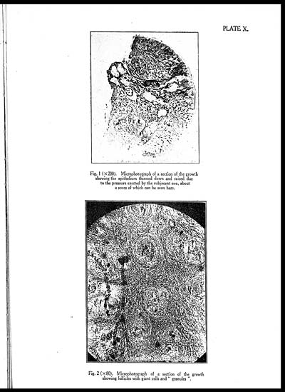
Fig. 1 (× 200). Microphotograph of a section of the growth
showing the epithelium thinned down and raised due
to the pressure exerted by the subjacent ova, about
a score of which can be seen here.
[NLS note: a graphic appears here - see image of page]
Fig. 2(× 80). Microphotograph of a section of the growth
showing follicles with giant cells and " granules "
Set display mode to: Large image | Zoom image | Transcription
Images and transcriptions on this page, including medium image downloads, may be used under the Creative Commons Attribution 4.0 International Licence unless otherwise stated. ![]()
| Permanent URL | https://digital.nls.uk/75227664 |
|---|---|
| Description | Covers articles from 1932. |
|---|


