

Medicine - Veterinary > Veterinary colleges and laboratories > Indian journal of veterinary science and animal husbandry > Volume 2, 1932 > Original articles > Etiology of bovine nasal granuloma
(154) Page 134
Download files
Individual page:
Thumbnail gallery: Grid view | List view
134 THE INDIAN JOURNAL OF VETERINARY SCIENCE AND ANIMAL HUSBANDRY [ II, II
may be short or long, slender throughout the length or broad at the base (c) an
ovoid—elongated egg with double terminal spine ( d) an egg of the " Spindalis "
type (Pl. IX, fig. 2.), as first described in India by Montgomery [1906] and also
found by Vriberg [1906] in Sumatra. The exact significance of so many types of
eggs is not clear.
Young, developing adult worms were also seen in the nasal discharge. The
young males are short and tuberculated with a prominent ventral sucker, which in
most cases being of yellow colour makes detection easy. The ventral sucker
approaches the anterior end of the worm, as it develops. The young females are
rather long and filiform.
Adult Schistosomes as seen in Pl. IX, fig. 3, have been removed at Muktesar
from the diseased tissue. This was removed surgically by slitting open the nose,
and deep sections were removed from the most posterior portion of the growths as
they were most likely to contain the healthy adults. An adult male was first isolat-
ed from the blood and other fluids collected during the surgical operation.
PATHOLOGICAL HISTOLOGY.
The material for histological study was obtained from specimens, which had
been collected at this Institute from about 12 cases of the disease from Assam,
Bombay, Mysore, Bihar and the Central Provinces from 1922 onwards.
Although other forms of bilharziosis, such as urinary and intestinal, are fre-
quently associated with a tendency towards malignancy and the formation of
polypoid growths, the essential histology of Bovine Nasal Granuloma is simply that
of granulation tissue, the exact character of which depends upon the degree and the
duration of the infection. An early growth is made up of very vascular young
connective tissue, fibroblasts in activity being in evidence. Old tumours are not so
highly vascular and contain a considerable amount of well formed fibrous tissue.
Eosinophilia, which is a notable feature of Nasal Granuloma, may be localised in the
periphery of the follicles, to be described later, or may even be diffusely scattered.
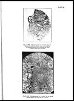
The epithelial covering of the growth is nearly completely intact excepting for
areas showing ulceration. The epithelium covering active lesions shows inflamma-
tory softening and may be actually thinned down due to the pressure exerted by the
ova attempting to work their way out. (Pl. ×, fig. 1.) Occasionally the apices of
the epithelial papillæ are found infected and may show degenerative changes. It is
primarily in the submucosa that the ova are deposited in quite appreciable numbers,
determining the nodular vascular masses of simple elevations of the mucosa which
characterise the disease. The submucous connective tissue is rather loosely dis-
posed and may show channel formations.
Set display mode to: Large image | Zoom image | Transcription
Images and transcriptions on this page, including medium image downloads, may be used under the Creative Commons Attribution 4.0 International Licence unless otherwise stated. ![]()
| Permanent URL | https://digital.nls.uk/75227661 |
|---|
| Description | Covers articles from 1932. |
|---|
| Description | Volumes 1-29 cover a wide range of topics related to veterinary science and animal husbandry. Black and white and colour plates accompany some articles. Each volume covers one year and has indexes to both authors and subjects at the end. Reviews, obituaries and abstracts are also included. |
|---|---|
| Shelfmark | IP/RA.11 |
| Additional NLS resources: | |
| Attribution and copyright: |
|
| Description | Reports from veterinary colleges and annual reports of the Imperial Bacteriologist. Plus volumes of the Indian Journal of Veterinary Science and Animal Husbandry covering 1931-1959. |
|---|---|
| Shelfmark | India Papers |
| Description | The Veterinary collection consists of 146 volumes dating from 1864 to 1959. Divided into veterinary diseases, colleges and laboratories and Civil Veterinary Departments. Extensive research on trypanosomiasis and rinderpest. Reports show how veterinary medicine controlled disease, maintained livestock and alleviated famine. They explore its effect on military and local communities. |
|---|---|
| Shelfmark | India Papers |
| Description | The India Papers collection contains publications of the central (Imperial) Government and many Indian states. Most states came under British rule. Much of the collection dates from between the post-Mutiny re-organisation of the Indian Government and Indian Independence in 1947. Some items published in London by John Murray. |
|---|---|
| Shelfmark | India Papers |
| Additional NLS resources: | |