Medicine - Veterinary > Veterinary colleges and laboratories > Indian journal of veterinary science and animal husbandry > Volume 4, 1934 > Original articles > Comparative study of Schistosoma spindalis, Montgomery, 1906 and Schistosoma nasalis, N. Sp.
(8) Page 2
Download files
Individual page:
Thumbnail gallery: Grid view | List view

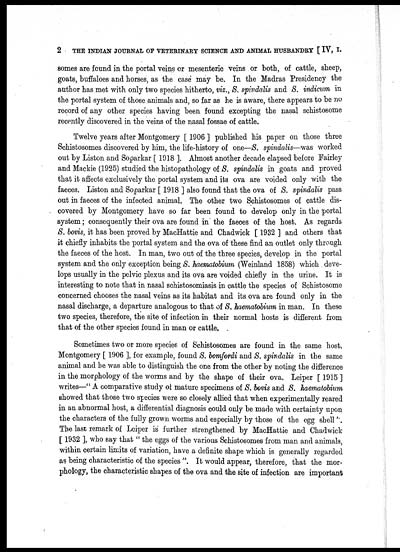
2 THE INDIAN JOURNAL OF VETERINARY SCIENCE AND ANIMAL HUSBANDRY [ IV, I.
somes are found in the portal veins or mesenteric veins or both, of cattle, sheep,
goats, buffaloes and horses, as the case may be. In the Madras Presidency the
author has met with only two species hitherto, viz., S. spindalis and S. indicum in
the portal system of those animals and, so far as he is aware, there appears to be no
record of any other species having been found excepting the nasal schistosome
recently discovered in the veins of the nasal fossae of cattle.
Twelve years after Montgomery [ 1906 ] published his paper on those three
Schistosomes discovered by him, the life-history of one— S. spindalis—was worked
out by Liston and Soparkar [ 1918 ]. Almost another decade elapsed before Fairley
and Mackie (1925) studied the histopathology of S. spindalis in goats and proved
that it affects exclusively the portal system and its ova are voided only with the
faeces. Liston and Soparkar [ 1918 ] also found that the ova of S. spindalis pass
out in faeces of the infected animal. The other two Schistosomes of cattle dis-
covered by Montgomery have so far been found to develop only in the portal
system; consequently their ova are found in the faeces of the host. As regards
S. bovis, it has been proved by MacHattie and Chadwick [ 1932 ] and others that
it chiefly inhabits the portal system and the ova of these find an outlet only through
the faeces of the host. In man, two out of the three species, develop in the portal
system and the only exception being S. haematobium (Weinland 1858) which deve-
lops usually in the pelvic plexus and its ova are voided chiefly in the urine. It is
interesting to note that in nasal schistosomiasis in cattle the species of Schistosome
concerned chooses the nasal veins as its habitat and its ova are found only in the
nasal discharge, a departure analogous to that of S. haematobium in man. In these
two species, therefore, the site of infection in their normal hosts is different from
that of the other species found in man or cattle.
Sometimes two or more species of Schistosomes are found in the same host.
Montgomery [ 1906 ], for example, found S. bomfordi and S. spindalis in the same
animal and he was able to distinguish the one from the other by noting the difference
in the morphology of the worms and by the shape of their ova. Leiper [ 1915 ]
writes—" A comparative study of mature specimens of S. bovis and S. haematobium
showed that those two species were so closely allied that when experimentally reared
in an abnormal host, a differential diagnosis could only be made with certainty upon
the characters of the fully grown worms and especially by those of the egg shell'.
The last remark of Leiper is further strengthened by MacHattie and Chadwick
[ 1932 ], who say that " the eggs of the various Schistosomes from man and animals,
within certain limits of variation, have a definite shape which is generally regarded
as being characteristic of the species ". It would appear, therefore, that the mor-
phology, the characteristic shapes of the ova and the site of infection are important
Set display mode to: Large image | Zoom image | Transcription
Images and transcriptions on this page, including medium image downloads, may be used under the Creative Commons Attribution 4.0 International Licence unless otherwise stated. ![]()
| Permanent URL | https://digital.nls.uk/75232067 |
|---|
| Description | Covers articles from 1934. |
|---|


![[Page1]](https://deriv.nls.uk/dcn4/7523/75232066.4.jpg)
