

Medicine - Veterinary > Veterinary colleges and laboratories > Indian journal of veterinary science and animal husbandry > Volume 3, 1933 > Original articles > Schistosoma indicum, Montgomery, 1906, as the cause of a persitant debility in equines in India, with a description of the lesions
(32) Page 18
Download files
Individual page:
Thumbnail gallery: Grid view | List view
18 THE INDIAN JOURNAL OF VETERINARY SCIENCE AND ANIMAL HUSBANDRY [ III, I
seems that the majority are capillaries or venules which have been abnormally
dilated but these may include occasional dilated lymph spaces. Apart from certain
large sized vessels which reveal a marked thinning and disruption of the muscular
coat, the most interesting lesions are presented by the endothelium of the smaller
vessels. The lesions consist of varying degrees of exudation into the endothelial
lining so that a sector of the vessel may become swollen to form a blunt finger-like
process filling a major portion of the lumen, whereas the endothelium of the other
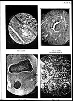
sectors is very severely mutilated or are even actually shed (Plate VI, Fig. 2).
Where the vessel is seen to contain a plug of inflammatory exudate, portions of the
desquamated endothelium may be recognisable as forming a part of it. With regard
to the probable mechanism by which the bloating of the endothelium takes place,
one notices polymorphonuclear leucocytes and masses of eosinophiles inside the
stumpy finger-like process. The fact that the immediate vicinity of the vessel wall
at the affected sector is heavily crowded with eosinophile cells suggests that a factor
such as an excessive lowering of blood pressure may have determined the endothelial
disruption, particularly when the parasitic toxin had already affected the susceptible
intima. Fairley [1920] has failed to confirm the finding of an endophlebitis of the
mesenteric veins as described by Letulle [1905] in his comprehensive article on
intestinal bliharziosis. The other prominent lesion of the blood vessels is represented
by degrees of endarteritis obliterans.
Apart from the pathological changes in the blood vessels supplying the intes-
tine, which have been referred to above, it must be remembered that the majority
of the nodular lesions in the submucosa or the subserosa originate in the embolic
eggmasses which form in the vessels of the place (Plate V, Fig. 2). It may be
mentioned here that the writer has encountered more than once up to 20 ova in a
single embolic focus in the intestine but although hundreds of liver sections like-
wise have been examined, he has never been able to find more than one ovum
in an individual nodule in the liver. Another remarkable fact is that in the
equine intestine, the large majority of Schistosome ova have been seen to be wholly
restricted to the layers of tissue situated below the mucosa (Plate II, Fig. 2),
whereas in the histological sections prepared from the intestine of natural cases of
S. indicum infection in the sheep and in the bull of both the plains and hill breeds,
the ova have been found almost exclusively in the mucous membrane with practi-
cally no tendency to nodule formation, the actual lesion being more cellular than
fibrous and not calcareous at all. Montgomery [1906] states that the large intes-
tine of equines is the only organ where ova of S. indicum could be found present
in any numbers. He found the ova in the mucosa only and thought that they must
occupy the submucous tissue or the capillary at some time.
Set display mode to: Large image | Zoom image | Transcription
Images and transcriptions on this page, including medium image downloads, may be used under the Creative Commons Attribution 4.0 International Licence unless otherwise stated. ![]()
| Permanent URL | https://digital.nls.uk/75229352 |
|---|
| Description | Covers articles from 1933. |
|---|
| Description | Volumes 1-29 cover a wide range of topics related to veterinary science and animal husbandry. Black and white and colour plates accompany some articles. Each volume covers one year and has indexes to both authors and subjects at the end. Reviews, obituaries and abstracts are also included. |
|---|---|
| Shelfmark | IP/RA.11 |
| Additional NLS resources: | |
| Attribution and copyright: |
|
| Description | Reports from veterinary colleges and annual reports of the Imperial Bacteriologist. Plus volumes of the Indian Journal of Veterinary Science and Animal Husbandry covering 1931-1959. |
|---|---|
| Shelfmark | India Papers |
| Description | The Veterinary collection consists of 146 volumes dating from 1864 to 1959. Divided into veterinary diseases, colleges and laboratories and Civil Veterinary Departments. Extensive research on trypanosomiasis and rinderpest. Reports show how veterinary medicine controlled disease, maintained livestock and alleviated famine. They explore its effect on military and local communities. |
|---|---|
| Shelfmark | India Papers |
| Description | The India Papers collection contains publications of the central (Imperial) Government and many Indian states. Most states came under British rule. Much of the collection dates from between the post-Mutiny re-organisation of the Indian Government and Indian Independence in 1947. Some items published in London by John Murray. |
|---|---|
| Shelfmark | India Papers |
| Additional NLS resources: | |