

Medicine - Veterinary > Veterinary colleges and laboratories > Indian journal of veterinary science and animal husbandry > Volume 3, 1933 > Original articles > Schistosoma indicum, Montgomery, 1906, as the cause of a persitant debility in equines in India, with a description of the lesions
(31) Page 17
Download files
Individual page:
Thumbnail gallery: Grid view | List view
SCHISTOSOMA INDICUM, MONTGOMERY, IN EQUINES 17
to chronic inflammatory changes may be so great that the bowel loses most of its
pliability and scarcely any light can pass through it. Individual pseudotubercles,
which may be situated anywhere between the mucous and peritoneal surfaces, show
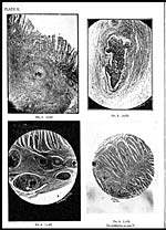
two or three sharply defined circles. (Plate II, Fig, 2). The outer circle is
fibrous and the central one contains a core of degenerated and calcareous material.
If a portion of the bowel showing early lesions is examined against a strong light,
one should be able to detect commencing vascular lesions from the centre of some of
which, the worm may be enucleated without much difficulty.
The mucous membrane of the intestine shows a marked mucoid degeneration
in a considerable portion of its length and this is associated with a profound infil-
tration with eosinophile leucocytes. It is strange that very few ova or none at all
are detected in the mucosa and from this, it seems probable that an extensive
diffusion of parasitic toxin must be responsible for these evenly distributed changes.
Small localised areas of inflammatory exudate or actual abscess formation may be
seen to have originated around half a dozen or more ova. The miracidium can be
seen inside the egg-shell which has undergone an irregular crenation. Actual
ulceration takes place by the desquamation of the epithelial cells, leading to the
formation of the peculiar minute ulcers described above and a varying amount of
blood pigment is deposited here and there. The muscular layer of the mucosa is in
tact practically throughout excepting for small gaps due to the dilation of the
arterioles which run between the mucosa and the submucosa (Plate VI, Fig. 1).
Actual breach of the muscularis mucosa takes place as a result of the disease process
advancing from the submucosa upwards and thus allowing the ova with the enclosed
maturing miracidia to be extruded into the outside world (Plate V, Fig. 1). As
has been seen before, the essential pathological lesions of the disease are found around
the ova. In early cases, it is the submucous coat only which provides the seat for the
production of the well known pseudotubercles [Fairley, 1920] and in an advanced
case, this layer is again the seat of the largest number of these nodular lesions. The
lesions extend to the subserosa (Plate V, Fig. 3) or in an extreme case may be
seen anywhere, but their formation is generally restricted to those situations which
have the most luxuriant blood supply. The nodules have been seen in between the
longitudinal layers of the muscular coat, these having been formed inside the
capillaries and venules which lie in strands of supporting intramuscular connective
tissue (Plate V, Fig. 4). The nodular lesions in the intestine follow the same
sequence of developmental stages as is seen in the case of similar lesions of the
liver. The blood vessels situated either in the submucosa or deeper are more or less
engorged and dilated. Now and again extremely dilated areas, rounded or oval in
shape, bounded by very thin walls and containing small round cells are seen im-
mediately below the muscularis mucosa. With regard to the origin of these, it
B 2
Set display mode to: Large image | Zoom image | Transcription
Images and transcriptions on this page, including medium image downloads, may be used under the Creative Commons Attribution 4.0 International Licence unless otherwise stated. ![]()
| Permanent URL | https://digital.nls.uk/75229349 |
|---|
| Description | Covers articles from 1933. |
|---|
| Description | Volumes 1-29 cover a wide range of topics related to veterinary science and animal husbandry. Black and white and colour plates accompany some articles. Each volume covers one year and has indexes to both authors and subjects at the end. Reviews, obituaries and abstracts are also included. |
|---|---|
| Shelfmark | IP/RA.11 |
| Additional NLS resources: | |
| Attribution and copyright: |
|
| Description | Reports from veterinary colleges and annual reports of the Imperial Bacteriologist. Plus volumes of the Indian Journal of Veterinary Science and Animal Husbandry covering 1931-1959. |
|---|---|
| Shelfmark | India Papers |
| Description | The Veterinary collection consists of 146 volumes dating from 1864 to 1959. Divided into veterinary diseases, colleges and laboratories and Civil Veterinary Departments. Extensive research on trypanosomiasis and rinderpest. Reports show how veterinary medicine controlled disease, maintained livestock and alleviated famine. They explore its effect on military and local communities. |
|---|---|
| Shelfmark | India Papers |
| Description | The India Papers collection contains publications of the central (Imperial) Government and many Indian states. Most states came under British rule. Much of the collection dates from between the post-Mutiny re-organisation of the Indian Government and Indian Independence in 1947. Some items published in London by John Murray. |
|---|---|
| Shelfmark | India Papers |
| Additional NLS resources: | |