Medicine - Institutions > Army health reports and medical documents > Scientific memoirs by medical officers of the Army of India > Part X, 1897 > 5 - On certain diseases of fungal and algal origin affecting economic plants in India
(151) Page 130
Thumbnail gallery: Grid view | List view

130
Scientific Memoirs by Medical Officers of the Army of India.
abnormal loss of water from the laminar surfaces. The excessive prevalence of
the blight may thus well be one factor in determining the very imperfect growth
of the Teak-tree in Lower Bengal.
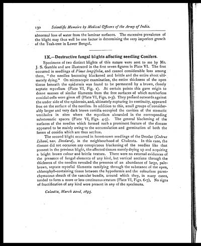
IX.—Destructive fungal blights affecting seedling Conifers.
Specimens of two distinct blights of this nature were sent to me by Mr.
J. S. Gamble and are illustrated in the first seven figures in Plate VI. The first
occurred in seedlings of Pinus longifolia, and caused considerable loss among
them, " the needles becoming blackened and brittle and the entire shoot ulti-
mately dying." On microscopic examination, the entire thickness of the open
tissue beneath the epidermis was found to be permeated by a brown, closely
septate mycelium (Plate VI, Fig. 1). At certain points this gave origin to
dense masses of similar filaments from the free surfaces of which numberless
conidial cells were given off (Plate VI, Figs. 2-3). They pressed outwards against
the under side of the epidermis, and, ultimately rupturing its continuity, appeared
free on the surface of the needles. In addition to this, small groups of consider-
ably larger and very dark brown conidia occupied the cavities of the stomatic
vestibules in sites where the mycelium abounded in the corresponding
substomatic spaces (Plate VI, Figs. 4-5). The general blackening of the
surfaces of the needles which formed such a prominent feature of the disease
appeared to be mainly owing to the accumulation and germination of both the
forms of conidia which are thus set free.
The second blight occurred in forest-sown seedlings of the Deodar (Cedrus
Libani, var. Deodara ), in the neighbourhood of Chakrata. In this case, the
disease did not occasion any conspicuous blackening of the needles like that
present in the previous blight, the affected tissues merely drying up and acquiring
a bright brown colour and brittle texture. There were no external evidences of
the presence of fungal elements of any kind, but vertical sections through the
thickness of the needles revealed the presence of an abundance of large, pale-
brown, septate mycelial filaments ramifying through the substance of the open,
chlorophyll-containing tissue between the hypodermis and the colourless paren-
chymatous sheath of the vascular bundle, around which they, in many cases,
tended to form a more or less continuous stratum (Plate VI, Figs. 6-7). No signs
of fructification of any kind were present in any of the specimens.
Calcutta, March 22nd, 1895.
Set display mode to: Large image | Zoom image | Transcription
Images and transcriptions on this page, including medium image downloads, may be used under the Creative Commons Attribution 4.0 International Licence unless otherwise stated. ![]()
| Permanent URL | https://digital.nls.uk/75003507 |
|---|
| Shelfmark | IP/QB.10 |
|---|---|
| Additional NLS resources: | |


