

Medicine - Institutions > Army health reports and medical documents > Scientific memoirs by medical officers of the Army of India > Part X, 1897 > 2 - Notes on the malarial parasite as observed in the blood during life, and in the tissues post-mortem, at Lahore, Punjab
(53) Page 47
Download files
Individual page:
Thumbnail gallery: Grid view | List view
Medical Officers of the Army of India.
47
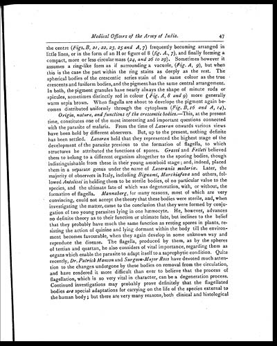
the centre (Figs. B, 21, 22, 23, 25 and A, 7 ) frequently becoming arranged in
little lines, or in the form of an H or figure of 8 (fig. A., 7 ), and finally forming a
compact, more or less circular mass (24, and 26 to 29 ). Sometimes however it
assumes a ring-like form as if surrounding a vacuole, (Fig. A, 9 ), but when
this is the case the part within the ring stains as deeply as the rest. The
spherical bodies of the crescentic series stain of the same colour as the true
crescents and fusiform bodies, and the pigment has the same central arrangement.
In both, the pigment granules have nearly always the shape of minute rods or
spicules sometimes distinctly red in colour ( Fig. A, 8 and 9 ) more generally
warm sepia brown. When flagella are about to develope the pigment again be-
comes distributed uniformly through, the cytoplasm (Fig. B, 16 and A, 14 ).
Origin, nature, and functions of the crescentic bodies. —This, at the present
time, constitutes one of the most interesting and important questions connected
with the parasite of malaria. From the time of Laveran onwards various views
have been held by different observers. But, up to the present, nothing definite
has been settled. Laveran held that they represented the highest stage of the
development of the parasite previous to the formation of flagella, to which
structures he attributed the functions of spores. Grassi and Felleti believed
them to belong to a different organism altogether to the sporing bodies, though
indistinguishable from these in their young amœboid stage; and, indeed, placed
them in a separate genus under the name of Laverania malariœ . Later, the
majority of observers in Italy, including Bignami, Marchiafava and others, fol-
lowed Antolisei in holding them to be sterile bodies, of no particular value to the
species, and the ultimate fate of which was degeneration, with, or without, the
formation of flagella. Mannaberg, for many reasons, most of which are very
convincing, could not accept the theory that these bodies were sterile, and, when
investigating the matter, came to the conclusion that they were formed by conju-
gation of two young parasites lying in one hæmocyte. He, however, advances
no definite theory as to their function or ultimate fate, but inclines to the belief
that they probably have much the same function as resting spores in plants, re-
sisting the action of quinine and lying dormant within the body till the environ-
ment becomes favourable, when they again develop in some unknown way and
reproduce the disease. The flagella, produced by them, as by the spheres
of tertian and quartan, he also considers of vital importance, regarding them as
organs which enable the parasite to adapt itself to a saprophytic condition. Quite
recently, Dr. Patrick Manson and Surgeon-Major Ross have devoted much atten-
tion to the changes undergone by these bodies on removal from the circulation,
and have rendered it more difficult than ever to believe that the process of
flagellation, which is so very vital in character, can be a degeneration process.
Continued investigations may probably prove definitely that the flagellated
bodies are special adaptations for carrying on the life of the species external to
the human body; but there are very many reasons, both clinical and histological
Set display mode to: Large image | Zoom image | Transcription
Images and transcriptions on this page, including medium image downloads, may be used under the Creative Commons Attribution 4.0 International Licence unless otherwise stated. ![]()
| Permanent URL | https://digital.nls.uk/75003201 |
|---|
| Shelfmark | IP/QB.10 |
|---|---|
| Additional NLS resources: | |
| Description | 12 volume series covers bacteriological research work from 1884-1901 by Indian Medical Service staff. Laboratory experiments are recorded in detail. Many are accompanied by illustrations, showing microscopic views and features of plant and animal matter. |
|---|---|
| Shelfmark | IP/QB.10 |
| Additional NLS resources: | |
| Description | 12 titles describe the health and sanitary conditions of the army in India. Lists British, European and Indian troops plus families of military personnel. 3 volumes of army regulations included. 73 volumes make up the Scientific Memoirs series, a journal by Indian Medical Service staff. |
|---|---|
| Description | The Institutions collection consists of 106 volumes from British India, dating from the 1860's to the 1940's. Divided into reports on medical institutions, army health, and lock hospitals. Education of indigenous medical students and maintenance of troop health shown in annual reports and statistical tables. |
|---|---|
| Description | The India Papers collection contains publications of the central (Imperial) Government and many Indian states. Most states came under British rule. Much of the collection dates from between the post-Mutiny re-organisation of the Indian Government and Indian Independence in 1947. Some items published in London by John Murray. |
|---|---|
| Shelfmark | India Papers |