Medicine - Veterinary > Civil Veterinary Departments > Civil Veterinary Department ledger series I-VI > Volume I - Rinderpest - cattle plague
(46) Page 18
Download files
Individual page:
Thumbnail gallery: Grid view | List view

18
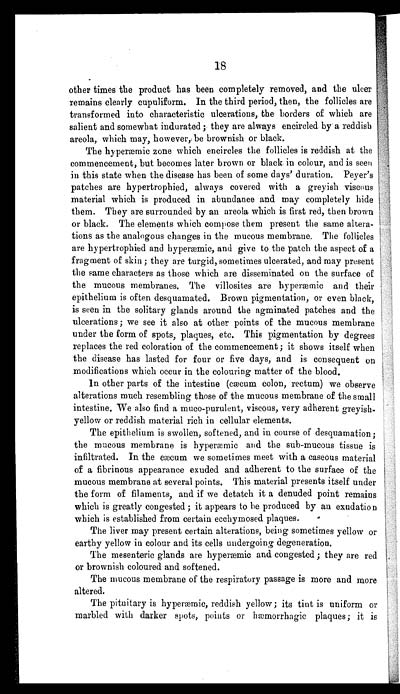
other times the product has been completely removed, and the ulcer
remains clearly cupuliform. In the third period, then, the follicles are
transformed into characteristic ulcerations, the borders of which are
salient and somewhat indurated ; they are always encircled by a reddish
areola, which may, however, be brownish or black.
The hyperæmic zone which encircles the follicles is reddish at the
commencement, but becomes later brown or black in colour, and is seen
in this state when the disease has been of some days' duration. Peyer's
patches are hypertrophied, always covered with a greyish viscous
material which is produced in abundance and may completely hide
them. They are surrounded by an areola which is first red, then brown
or black. The elements which compose them present the same altera-
tions as the analogous changes in the mucous membrane. The follicles
are hypertrophied and hyperæmic, and give to the patch the aspect of a
fragment of skin ; they are turgid, sometimes ulcerated, and may present
the same characters as those which are disseminated on the surface of
the mucous membranes. The villosites are hyperæmic and their
epithelium is often desquamated. Brown pigmentation, or even black,
is seen in the solitary glands around the agminated patches and the
ulcerations; we see it also at other points of the mucous membrane
under the form of spots, plaques, etc. This pigmentation by degrees
replaces the red coloration of the commencement; it shows itself when
the disease has lasted for four or five days, and is consequent on
modifications which occur in the colouring matter of the blood.
In other parts of the intestine (cæcum colon, rectum) we observe
alterations much resembling those of the mucous membrane of the small
intestine. We also find a muco-purulent, viscous, very adherent greyish-
yellow or reddish material rich in cellular elements.
The epithelium is swollen, softened, and in course of desquamation;
the mucous membrane is hyperæmic and the sub-mucous tissue is
infiltrated. In the cæcum we sometimes meet with a caseous material
of a fibrinous appearance exuded and adherent to the surface of the
mucous membrane at several points. This material presents itself under
the form of filaments, and if we detatch it a denuded point remains
which is greatly congested ; it appears to be produced by an exudation
which is established from certain ecchymosed plaques.
The liver may present certain alterations, being sometimes yellow or
earthy yellow in colour and its cells undergoing degeneration.
The mesenteric glands are hyperæmic and congested ; they are red
or brownish coloured and softened.
The mucous membrane of the respiratory passage is more and more
altered.
The pituitary is hyperæmic, reddish yellow ; its tint is uniform or
marbled with darker spots, points or hæmorrhagic plaques ; it is
Set display mode to: Large image | Zoom image | Transcription
Images and transcriptions on this page, including medium image downloads, may be used under the Creative Commons Attribution 4.0 International Licence unless otherwise stated. ![]()
| India Papers > Medicine - Veterinary > Civil Veterinary Departments > Civil Veterinary Department ledger series I-VI > Rinderpest - cattle plague > (46) Page 18 |
|---|
| Permanent URL | https://digital.nls.uk/75515902 |
|---|


