

Medicine - Veterinary > Veterinary colleges and laboratories > Indian journal of veterinary science and animal husbandry > Volume 13, 1943 > Original articles > Pleuropneumonia in goats with special reference to pasteuralla infection
(69) Page 47
Download files
Individual page:
Thumbnail gallery: Grid view | List view
P. G. PANDE 47
the help of a hand lens, appear to consist of minute granules varying in size
from a pin point to a pin head, which may be said to correspond with the lung
aveoli packed with red blood and other inflammatory cells.
The thoracic cavity contained a straw-coloured, or sometimes, a sero-
sanguineous fluid varying in amount from 500 c.c. to one litre. In cases
of longer duration the pleural membranes were found covered with fibrinous
deposits, and clots of fibrin were noticed enveloping the apical and the cardiac
lobes, particularly of the right lung. Another important and regular autopsy
finding was sero-sanguineous or fibrinous pericarditis.
Goats inoculated intravenously or subcutaneously with the culture or lung
emulsion showed petechial spots on all the serous membranes and the endothe-
lial lining of the heart, and areas of patchy congestion on the intestinal mucosa.
Histo-pathology
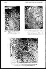
The lungs and the associated pleura were the only organs examined histo-
logically, as they were found chiefly affected. In cases of longer duration the
pleura was found thickly covered with a fibrinous exudate undergoing various
stages of organization. The inter-lobular septa in continuity with the thick-
ened pleura (Plate VI, fig. 1) contained leucocytic infiltration and engorged
capillaries. The lung tissue from a natural case showed extensive areas of grey
hepatization but areas of red hepatization were also discernible at places where
changes were of more recent duration, involving extension. In experimental
cases characteristic changes in their orthodox sequence were observed all in the
same lobe. The early microscopic lesion has been observed to begin with the
congestion of capillaries in the inter-alveolar septa, and the alveoli as a
sequence have been observed to contain (Plate VI, fig. 2) sero-cellular exudate
at this stage.
Areas of haemorrhagic infarcts, on section, showed thrombosis (Plate VI,
fig. 3) of minute bronchial vessels, engorgement of alveolar capillaries and
exudation of blood in the alveoli, along with other inflammatory cells.
Microscopical examination of stained slides by oil-immersion lens revealed a
great number of slender rods, 1 to 3 microns in length, seen with some difficulty
in densely packed alveoli, but more easily in less infiltrated areas where
they were numerous.
BACTERIOLOGY
Examination of smears from the cut surfaces of a consolidated portion
of the lung revealed a large number of gram-negative, slender and short rod
shaped bacilli, measuring from 1 to 3 microns in length, and having bipolar
appearance when stained with methylene blue or Leishman stain. They have
been seen to be most numerous in smears prepared from early lesions. The
same bipolar organisms could be readily and regularly obtained in pure culture
from the thöracic fluid, consolidated lung, pericardial fluid and the heart
blood. Cultures obtained from liver and kidney contained some contaminants,
Morphology. In. smears prepared from cultures grown on solid media.
the organism appears as short and plump rods, although few may appear as
elongated rods with slightly convex sides and rounded extremities. It is
gram-negative, non-motile, non-capsulated, non-sporulating and non-acid-fast
In smears prepared from broth culture the organism has been seen to occur
Set display mode to: Large image | Zoom image | Transcription
Images and transcriptions on this page, including medium image downloads, may be used under the Creative Commons Attribution 4.0 International Licence unless otherwise stated. ![]()
| Permanent URL | https://digital.nls.uk/75472530 |
|---|
| Description | Covers articles from 1943. |
|---|
| Description | Volumes 1-29 cover a wide range of topics related to veterinary science and animal husbandry. Black and white and colour plates accompany some articles. Each volume covers one year and has indexes to both authors and subjects at the end. Reviews, obituaries and abstracts are also included. |
|---|---|
| Shelfmark | IP/RA.11 |
| Additional NLS resources: | |
| Attribution and copyright: |
|
| Description | Reports from veterinary colleges and annual reports of the Imperial Bacteriologist. Plus volumes of the Indian Journal of Veterinary Science and Animal Husbandry covering 1931-1959. |
|---|---|
| Shelfmark | India Papers |
| Description | The Veterinary collection consists of 146 volumes dating from 1864 to 1959. Divided into veterinary diseases, colleges and laboratories and Civil Veterinary Departments. Extensive research on trypanosomiasis and rinderpest. Reports show how veterinary medicine controlled disease, maintained livestock and alleviated famine. They explore its effect on military and local communities. |
|---|---|
| Shelfmark | India Papers |
| Description | The India Papers collection contains publications of the central (Imperial) Government and many Indian states. Most states came under British rule. Much of the collection dates from between the post-Mutiny re-organisation of the Indian Government and Indian Independence in 1947. Some items published in London by John Murray. |
|---|---|
| Shelfmark | India Papers |
| Additional NLS resources: | |