

Medicine - Veterinary > Veterinary colleges and laboratories > Indian journal of veterinary science and animal husbandry > Volume 9, 1939 > Original articles > Observations on the existence of contagious bovine pleuro-pneumonia in British India, with an account of preliminary pathological investigation of cases of this disease reported from Assam
(19) Page 145
Download files
Individual page:
Thumbnail gallery: Grid view | List view

J. F. SHIRLAW 145
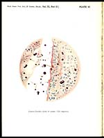
into the peribronchial interstitium (Plate VI) and their frequent numbers in
the blood vessels, correlated with the fairly frequent thrombosis of the
vessels, indicates a possible pathogenesis.
It is reasonable to consider that this case was one of mycotic pneumonia
but, as the tissues had been sent preserved in formalin, no further work was
possible to corroborate this opinion.
No detailed record of the clinical case was supplied with the material for
examination and no evidence could be obtained that the animal, from which
the lung tissues had been obtained, had died during the course of an epizootic.
Contagious bovine pleuro-pneumonia was again reported from Assam in
March, 1938.
With the dispatch of tissues and fluids for examination, the Disease In-
vestigation Officer, Assam, reported that these were selected from an animal
dying during the course of a severe epizootic in which there was considerable
mortality. " The disease appeared enzootic in particular localities, with a ten-
dency to affect fresh centres in the neighbourhood and was seasonal, occurring
during the advent of the monsoon. During the course of an outbreak, acute
and chronic cases are encountered, the former of four to six days and the latter
of twelve to twenty days duration. In acute cases, a moderate pyrexia of
104. 6°F. was noted and both lungs were equally involved. In chronic cases,
there was a marked tendency for only one lung to be affected. On 'post-
mortem examination, the lung from a chronic case was uniformly hepatized
and firmly adherent to the chest wall. The chest cavity contained about a
gallon of sanguineous fluid and a similar fluid was noticed on incision of the
lung tissue, the lobules of which appeared variegated ".
The tissues sent for examination were ampoules of heart blood, of lung
and of pleural exudate, and pieces of lung in 50 per cent glycerine and 5 per
cent formalin. Experimental transmission of the disease with the material
supplied was immediately instituted at Mukteswar.
No transmission experiments were conducted with the blood or with the
fluid exudates collected from the pleural cavity and the lung, which were
grossly contaminated. The material used for transmission experiments was a
portion of the glycerinated lung. This was washed in sterile distilled water
and finely triturated with sterile sand in normal saline. Two c. c. of the
supernatant fluid was inoculated subcutaneously into the rightside of the neck
of a hill bull, five years of age. A soft painful swelling, of the size of a golf ball,
developed within twenty-four hours, and persisted for sixteen days, when
it ruptured and discharged a thin pus from which no micro-organisms were
cultivable on selected media. The abscess healed uneventfully, with no ex-
tension, and during the entire period of observation extending over two months
no febrile or systemic disturbance was noted on the part of the animal. A
second hill bull of the same age, received a larger inoculation of five c. c. of the
saline suspension, with similar results.
B
Set display mode to: Large image | Zoom image | Transcription
Images and transcriptions on this page, including medium image downloads, may be used under the Creative Commons Attribution 4.0 International Licence unless otherwise stated. ![]()
| Permanent URL | https://digital.nls.uk/75246337 |
|---|
| Description | Covers articles from 1939. Please note that pagination starts at p.139 and plates at Plate V. |
|---|
| Description | Volumes 1-29 cover a wide range of topics related to veterinary science and animal husbandry. Black and white and colour plates accompany some articles. Each volume covers one year and has indexes to both authors and subjects at the end. Reviews, obituaries and abstracts are also included. |
|---|---|
| Shelfmark | IP/RA.11 |
| Additional NLS resources: | |
| Attribution and copyright: |
|
| Description | Reports from veterinary colleges and annual reports of the Imperial Bacteriologist. Plus volumes of the Indian Journal of Veterinary Science and Animal Husbandry covering 1931-1959. |
|---|---|
| Shelfmark | India Papers |
| Description | The Veterinary collection consists of 146 volumes dating from 1864 to 1959. Divided into veterinary diseases, colleges and laboratories and Civil Veterinary Departments. Extensive research on trypanosomiasis and rinderpest. Reports show how veterinary medicine controlled disease, maintained livestock and alleviated famine. They explore its effect on military and local communities. |
|---|---|
| Shelfmark | India Papers |
| Description | The India Papers collection contains publications of the central (Imperial) Government and many Indian states. Most states came under British rule. Much of the collection dates from between the post-Mutiny re-organisation of the Indian Government and Indian Independence in 1947. Some items published in London by John Murray. |
|---|---|
| Shelfmark | India Papers |
| Additional NLS resources: | |