

Medicine - Veterinary > Veterinary colleges and laboratories > Indian journal of veterinary science and animal husbandry > Volume 9, 1939 > Original articles > Cotugnia brotugerys, Meggitt, 1915 (from Gallus domesticus, Hosur Cattle Farm, Madras)
(241) Page 333
![[Page 334]](https://deriv.nls.uk/dcn4/7524/75247164.4.jpg)
Download files
Individual page:
Thumbnail gallery: Grid view | List view
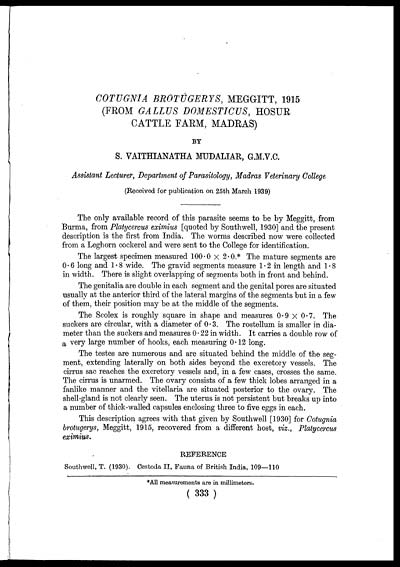
COTUGNIA BROTUGERYS, MEGGITT, 1915
(FROM GALLUS DOMESTICUS, HOSUR
CATTLE FARM, MADRAS)
BY
S. VAITHIANATHA MUDALIAR, G.M.V.C.
Assistant Lecturer, Department of Parasitology, Madras Veterinary College
(Received for publication on 25th March 1939)
The only available record of this parasite seems to be by Meggitt, from
Burma, from Platycercus eximius [quoted by Southwell, 1930] and the present
description is the first from India. The worms described now were collected
from a Leghorn cockerel and were sent to the College for identification.
The largest specimen measured 100.0 × 2.0.* The mature segments are
0.6 long and 1.8 wide. The gravid segments measure 1.2 in length and 1.8
in width. There is slight overlapping of segments both in front and behind.
The genitalia are double in each segment and the genital pores are situated
usually at the anterior third of the lateral margins of the segments but in a few
of them, their position may be at the middle of the segments.
The Scolex is roughly square in shape and measures 0.9 × 0.7. The
suckers are circular, with a diameter of 0.3. The rostellum is smaller in dia-
meter than the suckers and measures 0.22 in width. It carries a double row of
a very large number of hooks, each measuring 0.12 long.
The testes are numerous and are situated behind the middle of the seg-
ment, extending laterally on both sides beyond the excretory vessels. The
cirrus sac reaches the excretory vessels and, in a few cases, crosses the same.
The cirrus is unarmed. The ovary consists of a few thick lobes arranged in a
fanlike manner and the vitellaria are situated posterior to the ovary. The
shell-gland is not clearly seen. The uterus is not persistent but breaks up into
a number of thick-walled capsules enclosing three to five eggs in each.
This description agrees with that given by Southwell [1930] for Cotugnia
brotugerys, Meggitt, 1915, recovered from a different host, viz., Platycercus
eximius.
REFERENCE
Southwell, T. (1930). Cestoda II, Fauna of British India, 109—110
*All measurements are in millimeters.
(333)
Set display mode to: Large image | Zoom image | Transcription
Images and transcriptions on this page, including medium image downloads, may be used under the Creative Commons Attribution 4.0 International Licence unless otherwise stated. ![]()
| Permanent URL | https://digital.nls.uk/75247159 |
|---|
| Description | Covers articles from 1939. Please note that pagination starts at p.139 and plates at Plate V. |
|---|
| Description | Volumes 1-29 cover a wide range of topics related to veterinary science and animal husbandry. Black and white and colour plates accompany some articles. Each volume covers one year and has indexes to both authors and subjects at the end. Reviews, obituaries and abstracts are also included. |
|---|---|
| Shelfmark | IP/RA.11 |
| Additional NLS resources: | |
| Attribution and copyright: |
|
| Description | Reports from veterinary colleges and annual reports of the Imperial Bacteriologist. Plus volumes of the Indian Journal of Veterinary Science and Animal Husbandry covering 1931-1959. |
|---|---|
| Shelfmark | India Papers |
| Description | The Veterinary collection consists of 146 volumes dating from 1864 to 1959. Divided into veterinary diseases, colleges and laboratories and Civil Veterinary Departments. Extensive research on trypanosomiasis and rinderpest. Reports show how veterinary medicine controlled disease, maintained livestock and alleviated famine. They explore its effect on military and local communities. |
|---|---|
| Shelfmark | India Papers |
| Description | The India Papers collection contains publications of the central (Imperial) Government and many Indian states. Most states came under British rule. Much of the collection dates from between the post-Mutiny re-organisation of the Indian Government and Indian Independence in 1947. Some items published in London by John Murray. |
|---|---|
| Shelfmark | India Papers |
| Additional NLS resources: | |