

Medicine - Veterinary > Veterinary colleges and laboratories > Indian journal of veterinary science and animal husbandry > Volume 8, 1938 > Original articles > Studies on a natural outbreak of pigeon-pox
(227) Page 201
Download files
Individual page:
Thumbnail gallery: Grid view | List view
R. L. KAURA and S. GANAPATHY IYER 201
Material from the buccal lesions has not been used for subinoculation
purpose because it has been established in another paper by the authors [1936]
that in an identical condition in Indian fowls the causal agent of the buccal
diphtheritic lesions and the cutaneous lesions is one and the same filterable
virus.
TRANSMISSION EXPERIMENTS
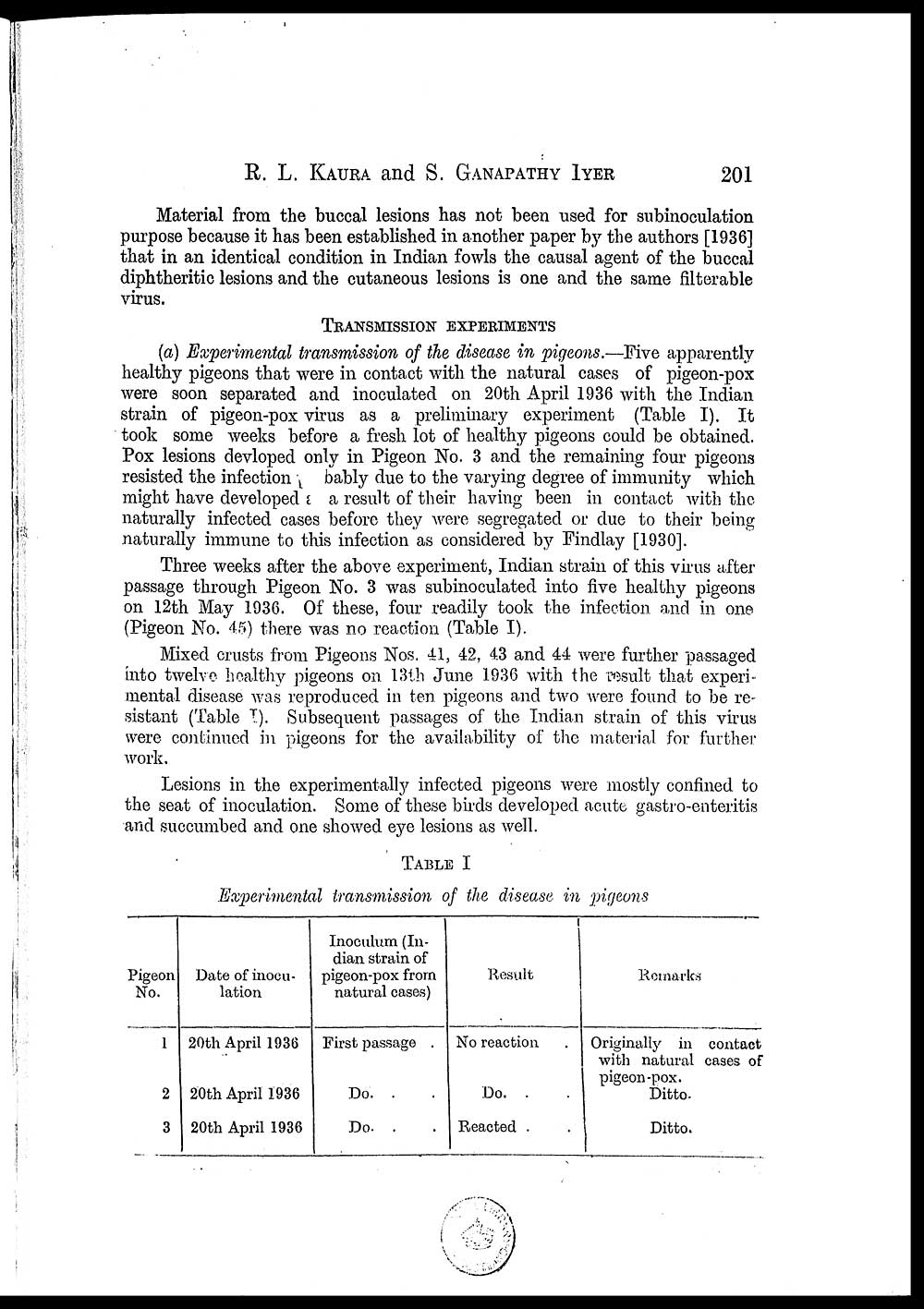
(a) Experimental transmission of the disease in pigeons.—Five apparently
healthy pigeons that were in contact with the natural cases of pigeon-pox
were soon separated and inoculated on 20th April 1936 with the Indian
strain of pigeon-pox virus as a preliminary experiment (Table I). It
took some weeks before a fresh lot of healthy pigeons could be obtained.
Pox lesions devloped only in Pigeon No. 3 and the remaining four pigeons
resisted the infection bably due to the varying degree of immunity which
might have developed area result of their having been in contact with the
naturally infected cases before they were segregated or due to their being
naturally immune to this infection as considered by Findlay [1930].
Three weeks after the above experiment, Indian strain of this virus after
passage through Pigeon No. 3 was subinoculated into five healthy pigeons
on 12th May 1936. Of these, four readily took the infection and in one
(Pigeon No. 45) there was no reaction (Table I).
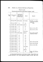
Mixed crusts from Pigeons Nos. 41, 42, 43 and 44 were further passaged
into twelve healthy pigeons on 13th June 1936 with the result that experi-
mental disease was reproduced in ten pigeons and two were found to be re-
sistant (Table I). Subsequent passages of the Indian strain of this virus
were continued in pigeons for the availability of the material for further
work.
Lesions in the experimentally infected pigeons were mostly confined to
the seat of inoculation. Some of these birds developed acute gastro-enteritis
and succumbed and one showed eye lesions as well.
TABLE I
Experimental transmission of the disease in pigeons
|
Pigeon No. |
Date of inocu- lation |
Inoculum (In- pigeon-pox from natural cases) |
Result |
Remarks |
|
1 |
20th April 1936 |
First passage . |
No reaction . |
Originally in contact |
|
2 |
20th April 1936 |
Do. . |
Do . . . |
Ditto. |
|
3 |
20th April 1936 |
Do. . |
Reacted . . |
Ditto. |
Set display mode to: Large image | Zoom image | Transcription
Images and transcriptions on this page, including medium image downloads, may be used under the Creative Commons Attribution 4.0 International Licence unless otherwise stated. ![]()
| Permanent URL | https://digital.nls.uk/75244189 |
|---|
| Description | Covers articles from 1938. |
|---|
| Description | Volumes 1-29 cover a wide range of topics related to veterinary science and animal husbandry. Black and white and colour plates accompany some articles. Each volume covers one year and has indexes to both authors and subjects at the end. Reviews, obituaries and abstracts are also included. |
|---|---|
| Shelfmark | IP/RA.11 |
| Additional NLS resources: | |
| Attribution and copyright: |
|
| Description | Reports from veterinary colleges and annual reports of the Imperial Bacteriologist. Plus volumes of the Indian Journal of Veterinary Science and Animal Husbandry covering 1931-1959. |
|---|---|
| Shelfmark | India Papers |
| Description | The Veterinary collection consists of 146 volumes dating from 1864 to 1959. Divided into veterinary diseases, colleges and laboratories and Civil Veterinary Departments. Extensive research on trypanosomiasis and rinderpest. Reports show how veterinary medicine controlled disease, maintained livestock and alleviated famine. They explore its effect on military and local communities. |
|---|---|
| Shelfmark | India Papers |
| Description | The India Papers collection contains publications of the central (Imperial) Government and many Indian states. Most states came under British rule. Much of the collection dates from between the post-Mutiny re-organisation of the Indian Government and Indian Independence in 1947. Some items published in London by John Murray. |
|---|---|
| Shelfmark | India Papers |
| Additional NLS resources: | |