

Medicine - Veterinary > Veterinary colleges and laboratories > Indian journal of veterinary science and animal husbandry > Volume 4, 1934 > Original articles > Comparative study of Schistosoma spindalis, Montgomery, 1906 and Schistosoma nasalis, N. Sp.
(24) Plate III
Download files
Individual page:
Thumbnail gallery: Grid view | List view
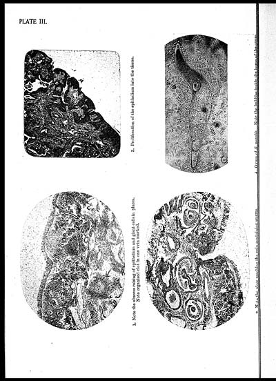
PLATE III.
[NLS note: a graphic appears here - see image of page]
1. Note the abscess raising of epithelium and giant cells in places.
Note organised clot in one vein marked.
[NLS note: a graphic appears here - see image of page]
2. Note the ulcer reaching the vein containing worms.
[NLS note: a graphic appears here - see image of page]
3. Proliferation of the epithelium into the tissue.
[NLS note: a graphic appears here - see image of page]
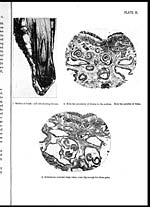
4. Oram of S. nasalis. Note the bubbles inside the horns of the ovum.
Set display mode to: Large image | Zoom image | Transcription
Images and transcriptions on this page, including medium image downloads, may be used under the Creative Commons Attribution 4.0 International Licence unless otherwise stated. ![]()
| Permanent URL | https://digital.nls.uk/75232115 |
|---|---|
| Description | Covers articles from 1934. |
|---|
| Description | Volumes 1-29 cover a wide range of topics related to veterinary science and animal husbandry. Black and white and colour plates accompany some articles. Each volume covers one year and has indexes to both authors and subjects at the end. Reviews, obituaries and abstracts are also included. |
|---|---|
| Shelfmark | IP/RA.11 |
| Additional NLS resources: | |
| Attribution and copyright: |
|
| Description | Reports from veterinary colleges and annual reports of the Imperial Bacteriologist. Plus volumes of the Indian Journal of Veterinary Science and Animal Husbandry covering 1931-1959. |
|---|---|
| Shelfmark | India Papers |
| Description | The Veterinary collection consists of 146 volumes dating from 1864 to 1959. Divided into veterinary diseases, colleges and laboratories and Civil Veterinary Departments. Extensive research on trypanosomiasis and rinderpest. Reports show how veterinary medicine controlled disease, maintained livestock and alleviated famine. They explore its effect on military and local communities. |
|---|---|
| Shelfmark | India Papers |
| Description | The India Papers collection contains publications of the central (Imperial) Government and many Indian states. Most states came under British rule. Much of the collection dates from between the post-Mutiny re-organisation of the Indian Government and Indian Independence in 1947. Some items published in London by John Murray. |
|---|---|
| Shelfmark | India Papers |
| Additional NLS resources: | |