

Medicine - Veterinary > Veterinary colleges and laboratories > Indian journal of veterinary science and animal husbandry > Volume 3, 1933 > Original articles > Etiology of bursati
(294) Page 236
Download files
Individual page:
Thumbnail gallery: Grid view | List view
236 THE INDIAN JOURNAL OF VETERINARY SCIENCE AND ANIMAL HUSBANDRY [III, III.
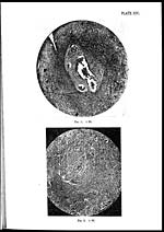
Plate XVIII, fig. 1.—The essential structure of an elongated kunkur, partially detached from
the mother tissue is to be seen. Eosinophil leucocytes are the most predominant cells
in this growth. Longitudinal section of the worm larva, with its transverse striation
can be seen occupying the blood vessel, which forms the nucleus for the production of
this kunkur.
Fig. 2.—Section of a bursati growth, showing a healthy worm in a healthy vessel. Only the
perivascular lymphocytic reaction is in evidence here. This stage is followed successively
by the stages represented by PI. XIX, fig. 1 and PI. XVIII, fig. 1.
Plate XIX, fig. 1.—Longitudinal section of a larva lying free in a blood vessel. Diffusive
action of the toxin can be judged by the homogeneous appearance of the border around
the vessel.
Fig. 2.—Photograph of a Habronema larva in a bursati section. The posterior end of the worm
is directed to the right, the anus being easily visible.
Fig. 3.—Photograph of the anterior end of a Habronema larva, obtained from a bursati section.
The cylindrical vestibule is clearly seen.
Set display mode to: Large image | Zoom image | Transcription
Images and transcriptions on this page, including medium image downloads, may be used under the Creative Commons Attribution 4.0 International Licence unless otherwise stated. ![]()
| Permanent URL | https://digital.nls.uk/75230356 |
|---|
| Description | Covers articles from 1933. |
|---|
| Description | Volumes 1-29 cover a wide range of topics related to veterinary science and animal husbandry. Black and white and colour plates accompany some articles. Each volume covers one year and has indexes to both authors and subjects at the end. Reviews, obituaries and abstracts are also included. |
|---|---|
| Shelfmark | IP/RA.11 |
| Additional NLS resources: | |
| Attribution and copyright: |
|
| Description | Reports from veterinary colleges and annual reports of the Imperial Bacteriologist. Plus volumes of the Indian Journal of Veterinary Science and Animal Husbandry covering 1931-1959. |
|---|---|
| Shelfmark | India Papers |
| Description | The Veterinary collection consists of 146 volumes dating from 1864 to 1959. Divided into veterinary diseases, colleges and laboratories and Civil Veterinary Departments. Extensive research on trypanosomiasis and rinderpest. Reports show how veterinary medicine controlled disease, maintained livestock and alleviated famine. They explore its effect on military and local communities. |
|---|---|
| Shelfmark | India Papers |
| Description | The India Papers collection contains publications of the central (Imperial) Government and many Indian states. Most states came under British rule. Much of the collection dates from between the post-Mutiny re-organisation of the Indian Government and Indian Independence in 1947. Some items published in London by John Murray. |
|---|---|
| Shelfmark | India Papers |
| Additional NLS resources: | |