16 THE INDIAN JOURNAL OF VETERINARY SCIENCE AND ANIMAL HUSBANDRY [III, I
particularly free from the lesions (Plate II, Fig. 2), though in very early cases
the venules in the submucous coat are the only seats where the pseudotubercles are
produced. In so far as the stomach is concerned, one must remember that certain
species of Habronema worms are quite frequently encountered there either lying
free on areas of superficial ulceration or embedded in fistulous tumours. As has
been seen already, the initial lesions start in the submucous bed of blood vessels of
the large intestines and it is the distal portions, namely, the large colon and the
rectum which reveal the earliest gross lesions, which again may easily be overlooked
unless one is particularly careful. Unless one is familiar with the minor lesions,
commonly associated with the early stages of a bilharzial enteritis, such as slight
congestion or petechial hæmorrhage, the proper significance of the picture may not
be understood. The intestinal contents may be semisolid or soft, coated with mucus
and slightly streaked with blood. As with other diseased conditions the character
and the extent of the lesions depend upon the duration and the severity of the
infection but neoplastic growths and tendency to malignancy, as are commonly
associated with certain forms of intestinal bilharziosis, are not seen in this equine
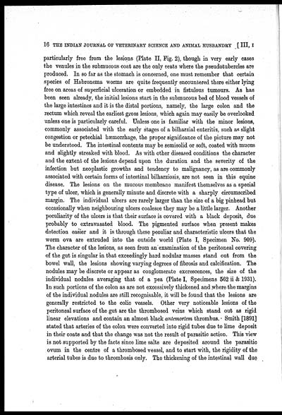
disease. The lesions on the mucous membrane manifest themselves as a special
type of ulcer, which is generally minute and discrete with a sharply circumscribed
margin. The individual ulcers are rarely larger than the size of a big pinhead but
occasionally when neighbouring ulcers coalesce they may be a little larger. Another
peculiarity of the ulcers is that their surface is covered with a black deposit, due
probably to extravasated blood. The pigmented surface when present makes
detection easier and it is through these peculiar and characteristic ulcers that the
worm ova are extruded into the outside world (Plate I, Specimen No. 909),
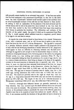
The character of the lesions, as seen from an examination of the peritoneal covering
of the gut is singular in that exceedingly hard nodular masses stand out from the
bowel wall, the lesions showing varying degrees of fibrosis and calcification. The
nodules may be discrete or appear as conglomerate excrescences, the size of the
individual nodules averaging that of a pea (Plate I, Specimens 562 ii & 1931).
In such portions of the colon as are not excessively thickened and where the margins
of the individual nodules are still recognisable, it will be found that the lesions are
generally restricted to the colic vessels. Other very noticeable lesions of the
peritoneal surface of the gut are the thrombosed veins which stand out as rigid
linear elevations and contain an almost black antemortem thrombus. Smith [1891]
stated that arteries of the colon were converted into rigid tubes due to lime deposit
in their coats and that the change was not the result of parasitic action. This view
is not supported by the facts since lime salts are deposited around the parasitic
ovum in the centre of a thrombosed vessel, and to start with, the rigidity of the
arterial tubes is due to thrombosis only. The thickening of the intestinal wall due


