

Medicine - Veterinary > Veterinary colleges and laboratories > Indian journal of veterinary science and animal husbandry > Volume 2, 1932 > Original articles > On the identity of the schistosome found in some cases of bovine nasal granuloma and some observations on a few other members of the schistosomidae
(409) Page 356
Download files
Individual page:
Thumbnail gallery: Grid view | List view
356 THE INDIAN JOURNAL OF VETERINARY SCIENCE AND ANIMAL HUSBANDRY [II, IV
Plate XXIX.—Schistosoma indicum.
Fig. 1. Anterior extremity of male.
Fig. 2. Female genitalia.
Figs. 3 & 4. Eggs from the intestine of hosts.
Plate XXX.—Schistosoma bovis.
Fig. 1. (a) Lateral view of the anterior extremity of male.
(b) A portion of male showing the division of the common intestinal
cæcum.
Fig. 2. Female genitalia.
Fig. 3. Uterine eggs.
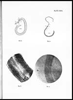
Plate XXXI.—Schistosoma turkestanicum.
Fig. 1. Entire male, lateral view.
Fig. 2. Entire female.
Plate XXXII.—
Fig. 1. A portion of male showing tuberculate nature of cuticle. ×128.
Fig. 2. A portion of male showing atuberculate nature of cuticle. × 33.
Fig. 3. Uterine egg, in-situ. ×830.
Explanation to Lettering.
du. eja., Ductus ejaculatorius.
e., Egg.
ex., Excretory bladder.
g.c., Gynæcophoric canal.
i.c., Intestinal cæcum.
oes., Œsophagus.
or. su., Oral sucker.
ov., Ovary.
sh. gl., Shell gland.
t., Testis.
ut., Uterus.
vent, su., Ventral sucker.
vcs. sem., Vesicula seminalis.
vit., Vitelline follicle.
Set display mode to: Large image | Zoom image | Transcription
Images and transcriptions on this page, including medium image downloads, may be used under the Creative Commons Attribution 4.0 International Licence unless otherwise stated. ![]()
| Permanent URL | https://digital.nls.uk/75228459 |
|---|
| Description | Covers articles from 1932. |
|---|
| Description | Volumes 1-29 cover a wide range of topics related to veterinary science and animal husbandry. Black and white and colour plates accompany some articles. Each volume covers one year and has indexes to both authors and subjects at the end. Reviews, obituaries and abstracts are also included. |
|---|---|
| Shelfmark | IP/RA.11 |
| Additional NLS resources: | |
| Attribution and copyright: |
|
| Description | Reports from veterinary colleges and annual reports of the Imperial Bacteriologist. Plus volumes of the Indian Journal of Veterinary Science and Animal Husbandry covering 1931-1959. |
|---|---|
| Shelfmark | India Papers |
| Description | The Veterinary collection consists of 146 volumes dating from 1864 to 1959. Divided into veterinary diseases, colleges and laboratories and Civil Veterinary Departments. Extensive research on trypanosomiasis and rinderpest. Reports show how veterinary medicine controlled disease, maintained livestock and alleviated famine. They explore its effect on military and local communities. |
|---|---|
| Shelfmark | India Papers |
| Description | The India Papers collection contains publications of the central (Imperial) Government and many Indian states. Most states came under British rule. Much of the collection dates from between the post-Mutiny re-organisation of the Indian Government and Indian Independence in 1947. Some items published in London by John Murray. |
|---|---|
| Shelfmark | India Papers |
| Additional NLS resources: | |