

Medicine - Veterinary > Veterinary colleges and laboratories > Indian journal of veterinary science and animal husbandry > Volume 2, 1932 > Original articles > Etiology of bovine nasal granuloma
(163) Page 137
Download files
Individual page:
Thumbnail gallery: Grid view | List view
ETIOLOGY OF BOVINE NASAL GRANULOMA 137
cells and their nuclei are pressed together into one or more solid masses and
take the stain deeply."
In the different parts of the matrix of the growth, empty spaces of various
shapes, rounded, oval, or spindle-shaped, are occasionally seen. These are
presumably a kind of mould left by the eggs. The resemblance of the shapes to
those of the actual ova discovered in the nasal discharges is worthy of note. The
matrix reveals groups of embedded eggs or empty irregular capsular shrunken shells.
Various stages of the embryo within the egg shell can be made out. Some appear
to be fully developed, other show a distinct segmenting embryo whilst some egg
shells are altogether collapsed and contain little or no organised material. Judging
by the appearance, shrunken and blackened shells with dark amorphous content
would appear to be eggs that have died due to unfavourable conditions. Inside
the shell of viable eggs, a distinct envelope (vitelline membrane) may be seen
enclosing the embryo. Some ova contain cells staining at the periphery, while
others show cells staining at the centre. In one ovum, at least, a fully matured,
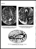
miracidium was seen still attached to the spiny portion of the egg shell (PI. XI,
fig. 2). In some sections, spaces which were originally occupied by the eggs are
seen occupied by mature or immature miracidia, a few of which are seen as leaf-
shaped bodies with an anterior cone, eosinophilic cephalic glands and a distinct
constriction between the cephalic and caudal regions. Some miracidia were
observed to have a blunt caudal extremity terminating abruptly, and these
presumably were the females.
In deeper sections, many coiled mucous glands and large blood vessels are
included. Some of the glands are apparently normal although containing some
discharge. Others may show hyperæmia. The glandular epithelium quite
frequently is noticed to undergo fatty degeneration and vacuolisation in many
places. In two samples of histological tissue, the discharges in the nasal gland
have been found to contain typical Bilharzia worms. Some glandular tubules were
definitely impacted with clusters of ova (Pl. XII, fig. 4), which would appear very
suitable to form the nucleus of "bilharzial pseudo-tubercles". It is difficult at
times to say definitely whether a pseudo-tubercle was initially formed by the
deposition of eggs in the tubules of nasal glands or within dilated capillaries or in
the matrix of the connective tissue itself.
Apart from the changes in the connective tissue of the nose described above,
the blood vessels show marked lesions in the shape of varying degrees of endar-
teritis, the lumen of some being altogether obliterated. The blood vessels in deeper
sections are generally larger and contain mature healthy worms, either singly or
in copula (Pl. XII, fig. 3), and may present considerable dilation and degrees of
Set display mode to: Large image | Zoom image | Transcription
Images and transcriptions on this page, including medium image downloads, may be used under the Creative Commons Attribution 4.0 International Licence unless otherwise stated. ![]()
| Permanent URL | https://digital.nls.uk/75227688 |
|---|
| Description | Covers articles from 1932. |
|---|
| Description | Volumes 1-29 cover a wide range of topics related to veterinary science and animal husbandry. Black and white and colour plates accompany some articles. Each volume covers one year and has indexes to both authors and subjects at the end. Reviews, obituaries and abstracts are also included. |
|---|---|
| Shelfmark | IP/RA.11 |
| Additional NLS resources: | |
| Attribution and copyright: |
|
| Description | Reports from veterinary colleges and annual reports of the Imperial Bacteriologist. Plus volumes of the Indian Journal of Veterinary Science and Animal Husbandry covering 1931-1959. |
|---|---|
| Shelfmark | India Papers |
| Description | The Veterinary collection consists of 146 volumes dating from 1864 to 1959. Divided into veterinary diseases, colleges and laboratories and Civil Veterinary Departments. Extensive research on trypanosomiasis and rinderpest. Reports show how veterinary medicine controlled disease, maintained livestock and alleviated famine. They explore its effect on military and local communities. |
|---|---|
| Shelfmark | India Papers |
| Description | The India Papers collection contains publications of the central (Imperial) Government and many Indian states. Most states came under British rule. Much of the collection dates from between the post-Mutiny re-organisation of the Indian Government and Indian Independence in 1947. Some items published in London by John Murray. |
|---|---|
| Shelfmark | India Papers |
| Additional NLS resources: | |