Medicine - Veterinary > Veterinary colleges and laboratories > Indian journal of veterinary science and animal husbandry > Volume 2, 1932 > Original articles > Rhinosporidiosis in equines
(60) Plate IV
Download files
Individual page:
Thumbnail gallery: Grid view | List view

PLATE IV.
[NLS note: a graphic appears here - see image of page]
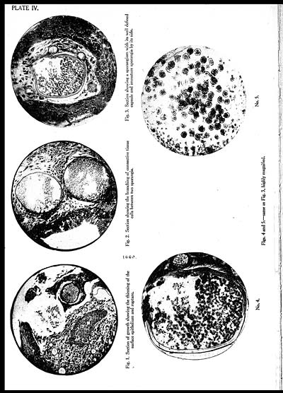
Fig. 1. Section of growth showing the thinning of the
surface epithelium and rupture.
[NLS note: a graphic appears here - see image of page]
Fig. 2. Section showing the branching of connective tissue
cells between two sporangia.
[NLS note: a graphic appears here - see image of page]
Fig. 3. Section showing a sporangium with its well defined
capsule and immature sporangia by its side.
[NLS note: a graphic appears here - see image of page]
No. 4.
[NLS note: a graphic appears here - see image of page]
No. 5.
Figs. 4 and 5.—same as Fig. 3, highly magnified.
Set display mode to: Large image | Zoom image | Transcription
Images and transcriptions on this page, including medium image downloads, may be used under the Creative Commons Attribution 4.0 International Licence unless otherwise stated. ![]()
| Permanent URL | https://digital.nls.uk/75227358 |
|---|---|
| Description | Covers articles from 1932. |
|---|


