Medicine - Institutions > Army health reports and medical documents > Scientific memoirs by officers of the Medical and Sanitary Departments of the Government of India > Number 50 - Preliminary report on an investigation into the etiology of Oriental sore in Cambay > Preliminary report on an investigation into the etiology of Oriental sore in Cambay
(22) Page 12
Download files
Individual page:
Thumbnail gallery: Grid view | List view

12
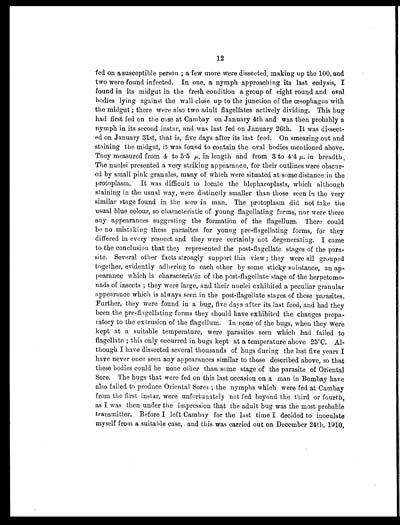
fed on a susceptible person; a few more were dissected, making up the 100, and
two were found infected. In one, a nymph approaching its last ecdysis, I
found in its midgut in the fresh condition a group of eight round and oval
bodies lying against the wall close up to the junction of the œsophagus with
the midgut; there were also two adult flagellates actively dividing. This bug
had first fed on the case at Cambay on January 4th and was then probably a
nymph in its second instar, and was last fed on January 26th. It was dissect-
ed on January 31st, that is, five days after its last feed. On smearing out and
staining the midgut, it was found to contain the oval bodies mentioned above.
They measured from 4 to 5.5 µ, in length and from 3 to 4.4 µ. in breadth.
The nuclei presented a very striking appearance, for their outlines were obscur-
ed by small pink granules, many of which were situated at some distance in the
protoplasm. It was difficult to locate the blepharoplasts, which although
staining in the usual way, were distinctly smaller than those seen in the very
similar stage found in the sore in man. The protoplasm did not take the
usual blue colour, so characteristic of young flagellating forms, nor were there
any appearances suggesting the formation of the flagellum. There could
be no mistaking these parasites for young pre-flagellating forms, for they
differed in every respect and they were certainly not degenerating. I came
to the conclusion that they represented the post-flagellate stages of the para-
site. Several other facts strongly support this view; they were all grouped
together, evidently adhering to each other by some sticky substance, an ap-
pearance which is characteristic of the post-flagellate stage of the herpetomo-
nads of insects; they were large, and their nuclei exhibited a peculiar granular
appearance which is always seen in the post-flagellate stages of these parasites.
Further, they were found in a bug, five days after its last feed, and had they
been the pre-flagellating forms they should have exhibited the changes prepa-
ratory to the extrusion of the flagellum. In none of the bugs, when they were
kept at a suitable temperature, were parasites seen which had failed to
flagellate; this only occurred in bugs kept at a temperature above 25°C. Al-
though I have dissected several thousands of bugs during the last five years I
have never once seen any appearances similar to those described above, so that
these bodies could be none other than some stage of the parasite of Oriental
Sore. The bugs that were fed on this last occasion on a man in Bombay have
also failed to produce Oriental Sores; the nymphs which were fed at Cambay
from the first instar, were unfortunately not fed beyond the third or fourth,
as I was then under the impression that the adult bug was the most probable
transmitter. Before I left Cambay for the last time I decided to inoculate
myself from a suitable case, and this was carried out on December 24th, 1910,
Set display mode to: Large image | Zoom image | Transcription
Images and transcriptions on this page, including medium image downloads, may be used under the Creative Commons Attribution 4.0 International Licence unless otherwise stated. ![]()
| Permanent URL | https://digital.nls.uk/75061485 |
|---|
| Shelfmark | IP/QB.10 |
|---|---|
| Additional NLS resources: | |


