Medicine - Institutions > Army health reports and medical documents > Scientific memoirs by officers of the Medical and Sanitary Departments of the Government of India > Number 18 - Haemogregarina gerbilli > Haemogregarina gerbilli
(18) Page 10
Download files
Individual page:
Thumbnail gallery: Grid view | List view

10
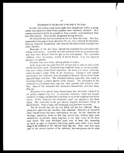
Development of the parasite in the body of the louse.
In order that feeding experiments might shew unequivocal results, a young
animal was selected in whose blood parasites were extremely abundant. As a
cursory examination of the fur revealed no lice, a number were transferred from
four older animals. They quickly disappeared among the hairs.
On the fourth day ova were noticed on the fur about the head. Two lice,
presumably belonging to those placed on the rat, were removed for dissection.
One was dissected immediately after removal, the other was left overnight in a
moist chamber.
Dissection of the first louse shewed very numerous free vermicules in the
mid-gut and intestine. A number were also scattered about the preparation, but
may have been derived from the gut as this was ruptured. The vermicules
exhibited active movements, mainly of lateral flexion. Very few encysted
parasites were present.
No forms were seen in the salivary glands or ovaries.
A dry preparation was made from the mid-gut and its contents and stained
with Romanowski's stain. Numerous large vermicule forms, in various attitudes,
were seen among altered blood corpuscles. As many as a dozen vermicules
were to be seen in single fields of the microscope. Compared with stained
specimens of free vermicules from the peripheral blood of the rat, these forms
appeared larger and finer. The chromatin masses, especially, were noted as
extending through a greater portion of the parasite. The protoplasm of the
vermicules was, in almost every case, free from granules and stained a clear
blue. Some of the vermicules had assumed a characteristic crescentic form
(Fig. 9.)
Dissection of the second louse shewed many free vermicules collected in
the pyloric ampulla (fig. 11). A motionless vermicule, which had become
crescentic in shape, was detected in the body cavity in the neighbourhood of the
rectum. In this dissection the alimentary canal was apparently free from
injury. The vermicules in the gut shewed sluggish movements chiefly of
lateral flexion. Only a single, still unchanged, encysted form was noted.
On the seventh day the rat was killed and the lice collected. Many
young lice, apparently just hatched, were observed. Dissection of several of
these shewed many very active vermicules in the gut. In the dissection of
the larger specimens, made on this day, certain very striking large cysts
undoubtedly of parasitic origin, lying free in the body cavity of the louse
were found. The most developed cysts were of large size, measuring
as much as 350µ in diameter, and being readily seen under a low power
lying in the abdomen of the infected louse (fig. 20). They were most often
seen in the anterior portion of the abdomen, but were present also in other
Set display mode to: Large image | Zoom image | Transcription
Images and transcriptions on this page, including medium image downloads, may be used under the Creative Commons Attribution 4.0 International Licence unless otherwise stated. ![]()
| Permanent URL | https://digital.nls.uk/75027427 |
|---|
| Shelfmark | IP/QB.10 |
|---|---|
| Additional NLS resources: | |


