

Medicine - Institutions > Army health reports and medical documents > Scientific memoirs by officers of the Medical and Sanitary Departments of the Government of India > Number 8 - Preliminary report on a parasite found in persons suffering from enlargement of the spleen in India > Preliminary report on a parasite found in persons suffering from enlargement of the spleen in India
(27) Page 17
Download files
Individual page:
Thumbnail gallery: Grid view | List view
17
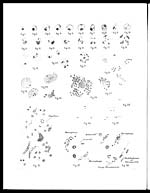
Explanation of Plate.
Fig. 1.—Small form of parasite without vacuole or secondary chromatin
body. Rarely seen.
Fig. 2.—Small form of parasite.
Fig. 3.—Large form of parasite.
Fig. 4.—Parasite showing rod shaped small chromatin mass.
Fig. 5.—Parasite showing small chromatin mass as a dot only.
Fig. 6.—Parasite showing " tail" joining chromatin masses.
Fig. 7.—Cockle shell shape with vacuole.
Fig. 8.— Ditto with, pink staining body substance and no vacuole.
Fig. 9.—Pear shaped form.
Fig. 10.—Form with faintly staining large chromatin mass.
Fig. 11.—Form showing bilobed chromatin mass.
Fig. 12.—Ditto heart-shaped chromatin mass.
Fig. 13.—Similar form to the last; seen in another direction.
Fig. 14.—Form with double large chromatin mass.
Fig. 15.—Longitudinal division into two forms.
Fig. 16.—Transverse division into two forms.
Fig. 17.—Developmental form showing the formation of 3 bodies.
Fig. 18.—Developmental form showing the formation of many bodies.
Fig. 19.—Forms apparently in altered red blood corpuscles.
Fig. 20.—Forms in bodies after treatment of blood with hypotonic ammo-
nium oxalate solution.
Fig. 21.—Pigment in apparent red cell.
Fig. 22.—Showing formation of apparent red cell bodies from macrophages
and leucocytes.
Fig. 23.—Parasites in polymorphonuclear leucocytes.
Fig. 24.—Parasite in a myelocyte.
Fig. 25.—Parasites in a large mononuclear leucocyte.
Fig. 26.—A macrophage containing the parasites.
Fig. 27.—A macrophage containing malarial pigment in addition to the
parasites.
Fig. 28.—Malignant tertian malaria. Young form showing two chromatin
dots.
Fig. 29.—Small forms of bovine piroplasma.
Fig. 30.—Large forms of bovine and canine piroplasma.
Fig. 31.—Section of liver stained by the modified Romanowsky method
showing macrophages in the liver capillaries.
Fig. 32.—Macrophages in the liver capillaries containing parasitic bodies.
Fig. 33.—Section of spleen showing the parasites in macrophages and in
large mononuclear cells; a parasite is seen in connection with an
endothelium or stroma cell.
Set display mode to: Large image | Zoom image | Transcription
Images and transcriptions on this page, including medium image downloads, may be used under the Creative Commons Attribution 4.0 International Licence unless otherwise stated. ![]()
| Permanent URL | https://digital.nls.uk/75026152 |
|---|---|
| Description | Explanation of plate |
| Shelfmark | IP/QB.10 |
|---|---|
| Additional NLS resources: | |
| Description | 61 volume new series covers bacteriological research work from 1902-1913 by Indian Medical Service staff. Laboratory experiments are recorded in detail. Many are accompanied by illustrations showing microscopic views and features of plant and animal matter. (Calcutta : Supt. of Govt. Print., India, 1885-1901.) |
|---|---|
| Shelfmark | IP/QB.10 |
| Description | 12 titles describe the health and sanitary conditions of the army in India. Lists British, European and Indian troops plus families of military personnel. 3 volumes of army regulations included. 73 volumes make up the Scientific Memoirs series, a journal by Indian Medical Service staff. |
|---|---|
| Description | The Institutions collection consists of 106 volumes from British India, dating from the 1860's to the 1940's. Divided into reports on medical institutions, army health, and lock hospitals. Education of indigenous medical students and maintenance of troop health shown in annual reports and statistical tables. |
|---|---|
| Description | The India Papers collection contains publications of the central (Imperial) Government and many Indian states. Most states came under British rule. Much of the collection dates from between the post-Mutiny re-organisation of the Indian Government and Indian Independence in 1947. Some items published in London by John Murray. |
|---|---|
| Shelfmark | India Papers |