

Medicine - Institutions > Army health reports and medical documents > Scientific memoirs by officers of the Medical and Sanitary Departments of the Government of India > Number 8 - Preliminary report on a parasite found in persons suffering from enlargement of the spleen in India > Preliminary report on a parasite found in persons suffering from enlargement of the spleen in India
(14) Page 8
Download files
Individual page:
Thumbnail gallery: Grid view | List view
8
end, and the small chromatin mass may often be seen undivided when the larger
masses are widely separated.
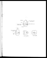
In the forms which apparently show transverse division the two large chro-
matin masses are at opposite poles separated by the two (or the as yet undivided)
small masses. Both these appearances represent the same process from different
points of view (see accompanying diagram). Division takes place in the line of
separation of what would be the two valves of the shell form. Seen from one
direction (A) the double pear-shaped form is seen. Seen from B. apparent
transverse division. The bilobed, heart-shaped and double forms of the chromatin
mass appear to be early stages of the same process.
In addition to the above, forms are seen showing division into three or more
bodies. These forms are more or less circular, of larger size than the largest of the.
single forms, and may be as large as a red blood corpuscle. In typical speci-
mens the large chromatin masses are arranged peripherally, whilst the small rod
shaped masses are nearer the centre. The formation of from 3 to 6 bodies is
most common.
Asexual and sexual forms.—No appearances have been seen by me which
seem to point to the presence of two forms of the parasite. Variations in the bodies
appear to be insignificant and to depend mostly upon the point of view from
which they are seen. The two most distinct forms are those with and those
without vacuoles. The forms showing uniform pink body substance are often
quite large. Another form is that with a very faintly staining chromatin mass.
Relation of the parasites to the red cell.—In ordinary preparations taken by
splenic puncture or in smears taken. post mortem, many of the bodies are. free
and found scattered over the slide. Other forms are seen included in leucocytes
and macrophages. Others again are found surrounded by some substance which
stains faintly blue and appears granular or reticular in structure. Bodies lying in
such material may occur singly or in numbers up to 10 or more. According to
Laveran this blue staining substance is the red cell rapidly disorganised by the
presence of the parasite. Ross mentions it only as a matrix.
Most of the apparently free bodies seen in films are, I believe, derived from
the leucocytes, and especially from enormous cells which are crowded with the
bodies and which readily become broken up. In many cases the bodies may be
seen to have been carried in streaks from such cells by the action of the slide or
needle used in spreading the film. Free bodies are generally scanty when blood
flows readily into the syringe during puncture. When this is the case very few
splenic cells are seen in the preparations and at the same time very few parasites.
They are more abundant when actual splenic pulp is used than when the films
are made from blood taken from the spleen. In sections enormous numbers of
Set display mode to: Large image | Zoom image | Transcription
Images and transcriptions on this page, including medium image downloads, may be used under the Creative Commons Attribution 4.0 International Licence unless otherwise stated. ![]()
| Permanent URL | https://digital.nls.uk/75026113 |
|---|
| Shelfmark | IP/QB.10 |
|---|---|
| Additional NLS resources: | |
| Description | 61 volume new series covers bacteriological research work from 1902-1913 by Indian Medical Service staff. Laboratory experiments are recorded in detail. Many are accompanied by illustrations showing microscopic views and features of plant and animal matter. (Calcutta : Supt. of Govt. Print., India, 1885-1901.) |
|---|---|
| Shelfmark | IP/QB.10 |
| Description | 12 titles describe the health and sanitary conditions of the army in India. Lists British, European and Indian troops plus families of military personnel. 3 volumes of army regulations included. 73 volumes make up the Scientific Memoirs series, a journal by Indian Medical Service staff. |
|---|---|
| Description | The Institutions collection consists of 106 volumes from British India, dating from the 1860's to the 1940's. Divided into reports on medical institutions, army health, and lock hospitals. Education of indigenous medical students and maintenance of troop health shown in annual reports and statistical tables. |
|---|---|
| Description | The India Papers collection contains publications of the central (Imperial) Government and many Indian states. Most states came under British rule. Much of the collection dates from between the post-Mutiny re-organisation of the Indian Government and Indian Independence in 1947. Some items published in London by John Murray. |
|---|---|
| Shelfmark | India Papers |