Medicine - Institutions > Army health reports and medical documents > Report of an investigation of the epidemic of malarial fever in Assam, or, kala-azar > Section V - Pathology and nature of kala-azar
(126) Page 101
Download files
Individual page:
Thumbnail gallery: Grid view | List view

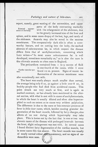
Pathology and nature of kála-ázar.
101
External appear-
ances of the body.
report, namely, great wasting of the extremities, and upper
parts of the body contrasting markedly
with the enlargement of the abdomen, due
to the greatly increased size of the liver and
spleen, and in some cases dropsy of the feet, legs, and rarely of
the abdomen. Anæmia may also be noted in the mucous
membranes. The comparatively dark skin is often a note-
worthy feature, and on cutting into the body, the marked
absence of subcutaneous fat, in which respect the disease
differs from that of anchylostomiasis, concerning which
Lutz writes—"In most cases subcutaneous fat is well
developed, sometimes remarkably so," as is also the case in
"the chlorotic anæmia so often seen in England.
Cardio vascular sys-
tem.
The pericardium contained from 1 to 5 ounces of fluid
in one-fourth of the cases, while I once
found 12 oz. present. Signs of recent in-
flammation of the serous membrane were
also occasionally met with.
The heart was nearly always much smaller than normal,
the average being only 6 oz., as against, a little over 8 oz., in
healthy people who had died from accidental causes. This
point struck me very much at first, and is again in
marked contrast, as has already been mentioned in the clini-
cal section, with what is met with in cases of anchylostomiasis,
in which the heart is usually dilated and often also hypertro-
phied to such an extent as to cause very evident palpitation.
The difference is due to the more or less constant presence of
fever in kála-ázar cases, which, besides causing granular de-
generation of the heart muscle, followed by fatty changes, also
allows of no rest during which hypertrophy may take
place. This is borne out by the fact that in one or two very
chronic cases of the disease with prolonged intervals between
the fever, slight enlargement was met with. In several cases
I found distinct fatty degeneration of the cardiac muscle, but
in some cases this was absent The heart muscle was usually
of nearly normal colour and consistency, and no signs of en-
docarditis were seen.
[NLS note: a graphic appears here - see image of 75014626.tif]
Set display mode to: Large image | Zoom image | Transcription
Images and transcriptions on this page, including medium image downloads, may be used under the Creative Commons Attribution 4.0 International Licence unless otherwise stated. ![]()
| Permanent URL | https://digital.nls.uk/75014624 |
|---|


