

Medicine - Institutions > Army health reports and medical documents > Scientific memoirs by medical officers of the Army of India > Part III, 1887 > 10 - Note on some aspects and relations of the blood-organisms in ague
(215) Transparency
Thumbnail gallery: Grid view | List view
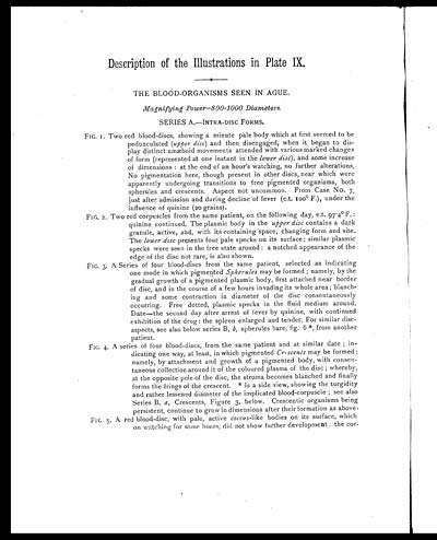
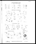
Description of the Illustrations in Plate IX.
THE BLOOD-ORGANISMS SEEN IN AGUE.
Magnifying Power—800–1000 Diameters.
SERIES A.—INTRA-DISC FORMS.
FIG.1. Two red blood-discs, showing a minute pale body which at first seemed to be
pedunculated (upper disc ) and then disengaged, when it began to dis-
play distinct amæboid movements attended with various marked changes
of form (represented at one instant in the lower disc ), and some increase
of dimensions: at the end of an hour’s watching, no further alterations.
No pigmentation here, though present in other discs, near which were
apparently undergoing transitions to free pigmented organisms, both
spherules and crescents. Aspect not uncommon. From Case No. 7,
just after admission and during decline of fever (e.t. 100° F.), under the
influence of quinine (20 grains).
FIG. 2. Two red corpuscles from the same patient, on the following day, e.t. 97.4° F.:
quinine continued. The plasmic body in the upper disc contains a dark
granule, active, and, with its containing space, changing form and site.
The lower disc presents four pale specks on its surface; similar plasmic
specks were seen in the free state around: a notched appearance of the
edge of the disc not rare, is also shown.
FIG. 3. A Series of four blood-discs from the same patient, selected as indicating
one mode in which pigmented Spherules may be formed; namely, by the
gradual growth of a pigmented plasmic body, first attached near border
of disc, and in the course of a few hours invading its whole area; blanch-
ing and some contraction in diameter of the disc consentaneously
occurring. Free dotted, plasmic specks in the fluid medium around.
Date—the second day after arrest of fever by quinine, with continued
exhibition of the drug: the spleen enlarged and tender. For similar disc-
aspects, see also below series B, b , spherules bare, fig. 6 *, from another
patient.
FIG. 4. A series of four blood-discs, from the same patient and at similar date; in-
dicating one way, at least, in which pigmented Crescents may be formed;
namely, by attachment and growth of a pigmented body, with consen-
taneous collection around it of the coloured plasma of the disc; whereby,
at the opposite pole of the disc, the stroma becomes blanched and finally
forms the fringe of the crescent. * Is a side view, showing the turgidity
and rather lessened diameter of the implicated blood-corpuscle; see also
Series B, a, Crescents, Figure 3, below. Crescentic organisms being
persistent, continue to grow in dimensions after their formation as above.
FIG. 5. A red blood-disc, with pale, active coccus -like bodies on its surface, which
on watching for some hours, did not show further development: the cor-
Set display mode to: Large image | Zoom image | Transcription
Images and transcriptions on this page, including medium image downloads, may be used under the Creative Commons Attribution 4.0 International Licence unless otherwise stated. ![]()
| Permanent URL | https://digital.nls.uk/75004657 |
|---|---|
| Description | Descriptions of the illustrations in plate IX |
| Shelfmark | IP/QB.10 |
|---|---|
| Additional NLS resources: | |
| Description | 12 volume series covers bacteriological research work from 1884-1901 by Indian Medical Service staff. Laboratory experiments are recorded in detail. Many are accompanied by illustrations, showing microscopic views and features of plant and animal matter. |
|---|---|
| Shelfmark | IP/QB.10 |
| Additional NLS resources: | |
| Description | 12 titles describe the health and sanitary conditions of the army in India. Lists British, European and Indian troops plus families of military personnel. 3 volumes of army regulations included. 73 volumes make up the Scientific Memoirs series, a journal by Indian Medical Service staff. |
|---|---|
| Description | The Institutions collection consists of 106 volumes from British India, dating from the 1860's to the 1940's. Divided into reports on medical institutions, army health, and lock hospitals. Education of indigenous medical students and maintenance of troop health shown in annual reports and statistical tables. |
|---|---|
| Description | The India Papers collection contains publications of the central (Imperial) Government and many Indian states. Most states came under British rule. Much of the collection dates from between the post-Mutiny re-organisation of the Indian Government and Indian Independence in 1947. Some items published in London by John Murray. |
|---|---|
| Shelfmark | India Papers |