Medicine - Institutions > Army health reports and medical documents > Scientific memoirs by medical officers of the Army of India > Part III, 1887 > 10 - Note on some aspects and relations of the blood-organisms in ague
(213) Transparency
‹‹‹ prev (212)
Reverse side of transparency
(214) next ›››
Reverse side of transparency
Download files
Individual page:
Thumbnail gallery: Grid view | List view

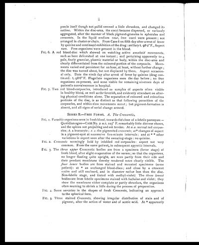
ii
puscle itself though not pallid seemed a little shrunken, and changed its
outline. Within the disc-area, the cocci became dispersed, or variously
aggregated, after the manner of black pigment-granules in spherules and
crescents. In the liquid medium near, free cocci were present; not
arranged in cluster or chain. From Case 6 on fifth day after arrest of fever
by quinine and continued exhibition of the drug: axillary t. 98.2° F., Aspect
rare. Free organisms were present in the blood.
FIG. 6. A red blood-disc which showed on watching active amæboid movements,
such as here delineated at one instant; and pertaining apparently to a
pale, finely granular, plasmic material or body, within the disc-area and
clearly differentiated from the coloured portion of the corpuscle. Move-
ments varied and persistent for an hour, at least, without further change;
the disc was turned about, but not displaced by them. Aspect occasion-
al only. Date the ninth day after arrest of fever by quinine (drug con-
tinued: t. 98.6° F. Flagellate organisms seen the day before; no free
organisms co-present, and none visible for remaining nineteen days of
patient’s convalescence in hospital.
FIG. 7. Two red blood-corpuscles, introduced as samples of aspects often visible
in healthy blood, as well as the feverish, and evidently attendant on alter-
ing physical conditions alone. The separation of coloured and colourless
portions of the disc, is as distinct as that following parasitism of the
corpuscles, and within slow movements occur; but pigment-formation is
absent, and all signs of serial change around.
SERIES B.—FREE FORMS. A. The Crescentic.
FIG. 1. Parasitic organisms seen in fresh blood, towards thé close of a febrile paroxysm —
Quotidian ague—CASE No. 2 m.t. 103° F. remarkably little distress shown,
and the spleen not projecting and not tender. At a. a. normal red corpus-
cles; b. a leucocyte; c. c. the pigmented crescents; at* changes of aspect
in a pigment-spot at successive five-minute intervals; and at * * other
variations in aspect seen after the sweating-stage: no quinine.
FIG. 2. Crescents seemingly held by infolded red corpuscles: aspect not very
common. From the same patient, in subsequent apyretic intervals.
FIG. 3. The three upper Crescentic bodies are from a specimen (fever stage) of
fresh blood, after slight evaporation of the serum; so that the organisms,
no longer floating quite upright, are seen partly from their side and
their pendant membrane thereby rendered more clearly visible. The
four lower bodies are from stained and mounted specimens (same
patient): at * an unchanged blood-discs; and close by a crescent
entire and still enclosed, and in diameter rather less than the disc.
Non-febrile stage, and tinted with methyl-violet: The three lowest
bodies are from febrile specimens stained with fuchsine and violet; they
show the membrane either complete or partly shrunken, the organisms
often seeming to shrink a little during the process of preparation.
FIG. 4. Some varieties in the shapes of fresh Crescents, indicating an approach
to the spherical form.
FIG. 5. Three stained Crescents, showing irregular distribution of stain and of
pigment, after the action of water and of acetic acid. At * apparently
Set display mode to: Large image | Zoom image | Transcription
Images and transcriptions on this page, including medium image downloads, may be used under the Creative Commons Attribution 4.0 International Licence unless otherwise stated. ![]()
| Permanent URL | https://digital.nls.uk/75004651 |
|---|
| Shelfmark | IP/QB.10 |
|---|---|
| Additional NLS resources: | |


