

Medicine - Institutions > Army health reports and medical documents > Scientific memoirs by medical officers of the Army of India > Part III, 1887 > 10 - Note on some aspects and relations of the blood-organisms in ague
(197) Transparency
Thumbnail gallery: Grid view | List view
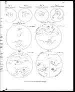
Description of the Illustrations.
PLATE V.
Figures 1 to 4 form a series, all magnified 500 diameters: Figures 1 and 2 are
hæmatophytes, and figures 3 and 4 are hæmatozoa, as described in the text.
The blood-disc introduced in each figure will serve to aid intercomparison of the
organisms.
FIGURE 1.—Bacillus anthracis (copied from Ziegler).
FIGURE 2.—Spirillum (Spirochæta) Obermeieri, aspects seen in blood during life;
straight and unfolded, also clustering and adhesion to red-discs
(human).
"3.—Tricho-monas sanguinis (rat) common aspects, when semi-quiescent.
"4.—Filaria sanguinis hominis (Lewis).
FIGURES 5 and 6.—Flagellated organisms from the rat's blood.
"5.—X 1000 diameters: (a ) from osmic acid preparations in which the uni-
lateral membrane is clearly indicated and a bright speck near the tail
ends. The blood-disc gives relative dimensions. (b ) from aniline-
stained preparations, in which the lighter stained edge corresponds
to the same lateral membrane; the flagellum is not wholly tinted (dated
Feb. 1885).
"6.— X 800 diameters. Changes undergone by rat-monads in fresh blood
diluted with 75 per cent. solution of common salt in water, hermetically
sealed and kept at 70° to 80°F. For description see the text: (a )
from a series of trials made in February 1885; (b ) from other experi-
ments made in September following, some slight difference being
apparent.
FIGURES 7 and 8.—Organisms from the blood of diseased animals affected with (surra).
" 7.— x 1000 diameters. From aniline-stained preparations: to the right are
four of the "monads " with two red discs (one shrivelled) and a larger
nucleated white cell; to the left are some smaller plasmic (?) particles*
x 1200 diameters as seen with 1/12 inch oil-immersion lens of Powell and
Lealand, London, in February 1885, and then designated as possibly
incipient flagellated organisms.
*They were sometimes found radiating round red-discs, or joined end to end, so as to appear of great
length, when their ends may unite; or joining laterally so as to seem thickened, doubled, or branching; or
clustering at various angles so as to form meshes or clumps; or intermingled variously. Below is seen the
attachment of the monads to red-discs, so striking in the fresh state.
FIGURE 8.— x 800 diameters. Organisms from inoculated animals, as named in the
figure. The differences apparent amongst the monads of mule, dog, and
monkey did not seem at all considerable, constant, or characteristic;
and it was considered that they hardly, if at all, surpassed a natural
variation, easily surmised and understood.
Set display mode to: Large image | Zoom image | Transcription
Images and transcriptions on this page, including medium image downloads, may be used under the Creative Commons Attribution 4.0 International Licence unless otherwise stated. ![]()
| Permanent URL | https://digital.nls.uk/75004597 |
|---|---|
| Description | Description of the illustrations. |
| Shelfmark | IP/QB.10 |
|---|---|
| Additional NLS resources: | |
| Description | 12 volume series covers bacteriological research work from 1884-1901 by Indian Medical Service staff. Laboratory experiments are recorded in detail. Many are accompanied by illustrations, showing microscopic views and features of plant and animal matter. |
|---|---|
| Shelfmark | IP/QB.10 |
| Additional NLS resources: | |
| Description | 12 titles describe the health and sanitary conditions of the army in India. Lists British, European and Indian troops plus families of military personnel. 3 volumes of army regulations included. 73 volumes make up the Scientific Memoirs series, a journal by Indian Medical Service staff. |
|---|---|
| Description | The Institutions collection consists of 106 volumes from British India, dating from the 1860's to the 1940's. Divided into reports on medical institutions, army health, and lock hospitals. Education of indigenous medical students and maintenance of troop health shown in annual reports and statistical tables. |
|---|---|
| Description | The India Papers collection contains publications of the central (Imperial) Government and many Indian states. Most states came under British rule. Much of the collection dates from between the post-Mutiny re-organisation of the Indian Government and Indian Independence in 1947. Some items published in London by John Murray. |
|---|---|
| Shelfmark | India Papers |