Medicine - Institutions > Army health reports and medical documents > Scientific memoirs by medical officers of the Army of India > Part III, 1887 > 10 - Note on some aspects and relations of the blood-organisms in ague
(152) Page 146
Download files
Individual page:
Thumbnail gallery: Grid view | List view

146
Scientific Memoirs by
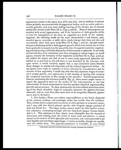
appearances similar to the above may all be met with; and, in addition, I noticed
others probably unconnected with the pigmented bodies, such as active pale coc-
cus-like granules, and even more striking heavings of the red-disc due to internal
amæba-like masses (vide Plate IX A, Figs. 5 and 6). None of these aspects are
attended with actual pigmentation, and if the formation of dark granules within
or from the hæmoglobin of the discs be regarded as a mark of the malaria-
organism, the following would be the more characteristic :—(a) minute, pale,
rounded spaces, vacuolate or solid, which slowly change their form and site, and
occasionally show dark spots (vide Plate IX A, Figs. 1 and 2); (b) pale-round-
ed spaces displaying within a dark pigment granule, which were seen in two or three
hours gradually to expand over the area of the disc, the pigment-particles augment-
ing, actively moving, and finally disposed as in free crescent or spheroid. In one field,
several red-discs were sometimes seen thus changing at various stages, as would
appear, towards the formation of free organisms (vide Plate IX A, Figs. 3 and 4):
not seldom the aspect was that of one or more small pigmented spherules im-
bedded in, or attached to, the red-discs, as was described by Dr. Laveran; and,
upon review, it would certainly appear that a real connection exists between
these changes in certain red-corpuscles and the isolated pigmented bodies. As
my figures were made at cessation of fever, followed by convalescence and no
increase of free organisms, I could only infer that they represent stages of arrest-
ed if normal growth; and opportunity is still wanting of tracing with certainty
the completed transition of disc-change to free parasite. 1Stained preparations,
however, confirming this inference (vide B, Fig. 3), there seems ample reason to
regard the pigmented spots on the surface of the red-corpuscles as not less
characteristic of blood-contamination than the manifest detached organisms they
precede and accompany. To show permanently the intra-cellular structures, stain-
ing of the blood should be slight or moderate, otherwise the pigment becomes
obscured; and as, even then, tints seem often to be irregularly disposed, appear-
ances may be deceptive.
b. Free bodies.— These are evident super-additions to the blood, dispersed
in the common medium, and seemingly rather less mobile than the red-discs.
They include distinct pigmented structures, of either globular or crescentic shape;
and I may add also dotted plasmic specks, with irregular opaque particles of
dark and bluish hue. The larger bodies attract notice by their hyaline colour-
less substance, contrasting strongly with the deep-hued opaque clumps imbed-
ded in it. This substance is firm, elastic, more refrangible than the material of
leucocytes, and resisting more the action of weak acids and alkalies; it seems
denser on the exterior of both spherules and crescents, and, continuous with it are
their flagellar and membrane appendages: it stains as readily, though not equally,
with the aniline dyes. The pigment-constituent is invariable, as very minute
1Subsequent consecutive blood-scrutinies ahve strongly indicated such transition.
Set display mode to: Large image | Zoom image | Transcription
Images and transcriptions on this page, including medium image downloads, may be used under the Creative Commons Attribution 4.0 International Licence unless otherwise stated. ![]()
| Permanent URL | https://digital.nls.uk/75004462 |
|---|
| Shelfmark | IP/QB.10 |
|---|---|
| Additional NLS resources: | |


