

Medicine - Institutions > Army health reports and medical documents > Scientific memoirs by medical officers of the Army of India > Part VII, 1892 > 3 - On nodular disease of the intestine in sheep
(66) Transparency
Thumbnail gallery: Grid view | List view

Explanation of Plate.
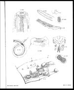
A FIG. 1. Œsophagostoma columbianum, Curtice.
Head x 280 diameters. The two padilke which serve as openings of
the lateral canals are seen on either side. Of the other four tubercles,
only one is shown, to avoid confusing the outlines of the armature.
" 2. Split preparation of the head of the same x 280 diameters showing, first, a
ring of plates with semilunar ends, next the circlet of fimbriæ, and
lastly, the circlet of bidentate teeth, below which is the wide œsophagus.
" 3. Bursa of male Œsophagostoma partially spread x 70 diameters.
" 4. Ovum of Œsophagostoma x 440 diameters taken from the clot adhering
to the vulva.
" 5. Trichonema stage of Œsophagostoma from tumour of intestine of sheep.
" 6. A portion of the large intestine of a sheep affected with nodular disease
of the intestine. Natural size.
B. Trichosomum verrucosum, sp. n.
" 7. Trichosomum verrucosum, ♀, natural size.
" 8. Head of the same x 28 diameters.
" 9. Caudal extremity of the same x 28 diameters—
a —anus.
v —vulva.
u —uterus.
i —intestine.
" 10. Ovum of above, from uterus x 440 diameters.
Set display mode to: Large image | Zoom image | Transcription
Images and transcriptions on this page, including medium image downloads, may be used under the Creative Commons Attribution 4.0 International Licence unless otherwise stated. ![]()
| Permanent URL | https://digital.nls.uk/75001345 |
|---|---|
| Description | Explanation of plate |
| Shelfmark | IP/QB.10 |
|---|---|
| Additional NLS resources: | |
| Description | 12 volume series covers bacteriological research work from 1884-1901 by Indian Medical Service staff. Laboratory experiments are recorded in detail. Many are accompanied by illustrations, showing microscopic views and features of plant and animal matter. |
|---|---|
| Shelfmark | IP/QB.10 |
| Additional NLS resources: | |
| Description | 12 titles describe the health and sanitary conditions of the army in India. Lists British, European and Indian troops plus families of military personnel. 3 volumes of army regulations included. 73 volumes make up the Scientific Memoirs series, a journal by Indian Medical Service staff. |
|---|---|
| Description | The Institutions collection consists of 106 volumes from British India, dating from the 1860's to the 1940's. Divided into reports on medical institutions, army health, and lock hospitals. Education of indigenous medical students and maintenance of troop health shown in annual reports and statistical tables. |
|---|---|
| Description | The India Papers collection contains publications of the central (Imperial) Government and many Indian states. Most states came under British rule. Much of the collection dates from between the post-Mutiny re-organisation of the Indian Government and Indian Independence in 1947. Some items published in London by John Murray. |
|---|---|
| Shelfmark | India Papers |