

Medicine - Institutions > Army health reports and medical documents > Scientific memoirs by medical officers of the Army of India > Part V, 1890 > 5 - On a Chrysomyxa on Rhododendron arboreum, Sm. (Chrysomyxa Himalense, Nov. Sp.)
(107) Transparency
Thumbnail gallery: Grid view | List view
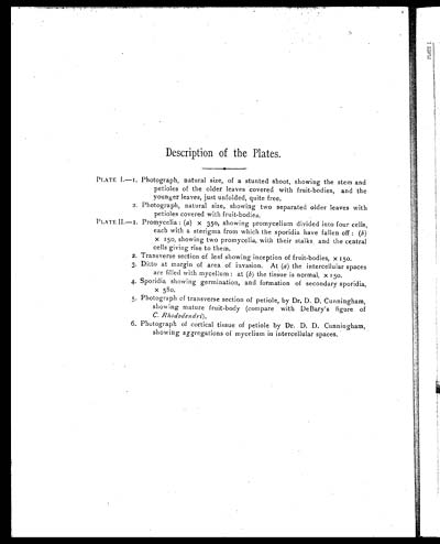
Description of the Plates.
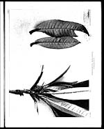
PLATE I.—1. Photograph, natural size, of a stunted shoot, showing the stem and
petioles of the older leaves covered with fruit-bodies, and the
younger leaves, just unfolded, quite free.
2. Photograph, natural size, showing two separated older leaves with
petioles covered with fruit-bodies.
PLATE II—1. Promycelia: (a ) x 350, showing promycelium divided into four cells,
each with a sterigma from which the sporidia have fallen off: (b )
x 150, showing two promycelia, with their stalks and the central
cells giving rise to them.
2. Transverse section of leaf showing inception of fruit-bodies, x 150.
3.Ditto at margin of area of invasion. At (a ) the intercelluiar spaces
are filled with mycelium: at (b ) the tissue is normal, x 150.
4. Sporidia showing germination, and formation of secondary sporidia,
x 580.
5. Photograph of transverse section of petiole, by Dr. D. D. Cunningham,
showing mature fruit-body (compare with DeBary's figure of
C. Rhododendri ).
6. Photograph of cortical tissue of petiole by Dr. D. D. Cunningham,
showing aggregations of mycelium in intercellular spaces.
Set display mode to: Large image | Zoom image | Transcription
Images and transcriptions on this page, including medium image downloads, may be used under the Creative Commons Attribution 4.0 International Licence unless otherwise stated. ![]()
| Permanent URL | https://digital.nls.uk/75000569 |
|---|---|
| Description | Description of the plates |
| Shelfmark | IP/QB.10 |
|---|---|
| Additional NLS resources: | |
| Description | 12 volume series covers bacteriological research work from 1884-1901 by Indian Medical Service staff. Laboratory experiments are recorded in detail. Many are accompanied by illustrations, showing microscopic views and features of plant and animal matter. |
|---|---|
| Shelfmark | IP/QB.10 |
| Additional NLS resources: | |
| Description | 12 titles describe the health and sanitary conditions of the army in India. Lists British, European and Indian troops plus families of military personnel. 3 volumes of army regulations included. 73 volumes make up the Scientific Memoirs series, a journal by Indian Medical Service staff. |
|---|---|
| Description | The Institutions collection consists of 106 volumes from British India, dating from the 1860's to the 1940's. Divided into reports on medical institutions, army health, and lock hospitals. Education of indigenous medical students and maintenance of troop health shown in annual reports and statistical tables. |
|---|---|
| Description | The India Papers collection contains publications of the central (Imperial) Government and many Indian states. Most states came under British rule. Much of the collection dates from between the post-Mutiny re-organisation of the Indian Government and Indian Independence in 1947. Some items published in London by John Murray. |
|---|---|
| Shelfmark | India Papers |