Medicine - Institutions > Army health reports and medical documents > Scientific memoirs by medical officers of the Army of India > Part IV, 1889 > 2 - Notes on the life-history of Ravenelia sessilis, B. and Ravenelia stictica, B. and Br.
(40) Page 34
Download files
Individual page:
Thumbnail gallery: Grid view | List view

34
Scientific Memoirs by
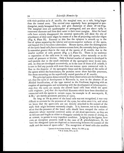
with their position as in R. sessilis, the marginal ones, as a rule, being larger
than the central ones. The central ones superiorly have pentagonal or qua-
drangular, rarely hexagonal faces, and give diameters of about 18.1x16.5µ.
The marginal ones are quadrangular or pentagonal, and are about 19.8µ in
transverse diameter and from their outer to their inner margins. After the basal
cells have entirely disappeared the external spore-cells still show the site of
attachment of their outer edges by means of a line of projecting cuticular ridges
(Fig. 6, Plate II). External to this line the epispore is smooth up to the
line of spines separating the inferior and extero-superior surfaces of the spore,
and beyond this it becomes tuberculate. Mature spores, after the disintegration
of the cystic basal cells, have a concavo-convex form, the convexity being relative-
ly somewhat greater than in the case of the spores of R. sessilis, due to the
smaller number of cells present (Fig. 5 b, Plate II). There is no tendency
to separation of the cells even in very old spores, either naturally or as the
result of pressure or friction. Only a limited number of spores is developed in
each pustule due to the small extension of the sporogenic issue in any case,
and, as they are developed successively, as in the case of those of R. sessilis, it
is rare to find any pustule with more than one mature spore connected with it.
Due to the deepsite of the sporogenic tissue and the inclusion of the stalks of
the spores within the host-tissues, the pustules never show hair-like processes,
like those occurring on the superficially seated pustules of R. sessilis.
The principal points demonstrated by these observations are the following:—
1st, that the cycle of development in Ravenelia is, save in the absence of an
Æcidial fructification, of the type normal to the Uredineæ, beginning with an
eruption of spermogonia and terminating with the development of teleutospores;
2nd, that the cysts are merely the altered basal cells from which the spore
cells originate; 3rd, that the mycelioid filaments which have been described as
connected with the spores in certain cases, for example in R. macrocystis, B
& Br.,1are merely persistent portions of cysts.
So long as the presence of the basal cells was not recognised, it was very
difficult to account for the presence of the cysts, but when once it is, and when
we know that the spore-cells are not directly attached to the summit of the
stalk their origin becomes extremely simple. It is, however, not to be wonder-
ed at that the presence of the basal cells should have escaped the notice of
observers whose material consisted solely of old dried specimens, as they are
so delicate and fragile as either to disappear entirely in the course of drying, or,
at utmost, to persist in very imperfect condition. Judging by the figures, how-
ever, an exception presents itself in the case of R. glandulœformis, B., for
here the elongated cysts are depicted as intervening between the summit of the
stalk and the mass of spore-cells.2The recognition of basal cells also enables
1Journal of the Linnean Society: Botany, Vol. XIV, p. 93.
2M. C. Cooke, M.A., op. cit,
Set display mode to: Large image | Zoom image | Transcription
Images and transcriptions on this page, including medium image downloads, may be used under the Creative Commons Attribution 4.0 International Licence unless otherwise stated. ![]()
| Permanent URL | https://digital.nls.uk/75000000 |
|---|
| Shelfmark | IP/QB.10 |
|---|---|
| Additional NLS resources: | |


