

Medicine - Disease > Report on the cultivation of protesoma, Labbé, in grey mosquitoes
(29) Page 21
![Page [22]](https://deriv.nls.uk/dcn4/7444/74448043.4.jpg)
Download files
Individual page:
Thumbnail gallery: Grid view | List view
21
APPENDIX.
Description of the Grey Mosquitos.
The following description of the mosquitos which carry proteosoma, and which I call "grey"
mosquitos, has been very kindly furnished me by G. C. Dudgeon, Esq., of Pankabari, whose ento-
mological experience is well known:-
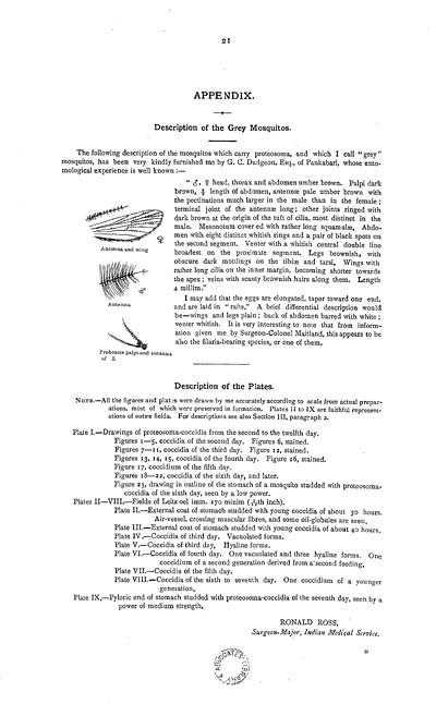
" ?, ? head, thorax and abdomen umber brown. Palpi dark
brown, 2/3 length of abdomen, antenn pale umber brown with
the pectinations much larger in the male than in the female;
terminal joint of the antenn long; other joints ringed with
dark brown at the origin of the tuft of cilia, most distinct in the
male. Mesonotum cover ed with rather long squamul. Abdo-
men with eight distinct whitish rings and a pair of black spots on
the second segment. Venter with a whitish central double line
broadest on the proximate segment. Legs brownish, with
obscure dark mottlings on the tibi and tarsi. Wings with
rather long cilia on the inner margin, becoming shorter towards
the apex; veins with scanty brownish hairs along them. Length
4 millim."
I may add that the eggs are elongated, taper toward one end,
and are laid in "rafts." A brief differential description would
be-wings and legs plain; back of abdomen barred with white;
venter whitish. It is very interesting to note that from inform-
ation given me by Surgeon-Colonel Maitland, this appears to be
also the filaria-bearing species, or one of them.
Description of the Plates.
NOTE.-All the figures and plates were drawn by me accurately according to scale from actual prepar-
ations, most of which were preserved in formation. Plates II to IX are faithful represent-
ations of entire fields. For descriptions see also Section III, paragraph 2.
Plate I.-Drawings of proteosoma-coccidia from the second to the twelfth day.
Figures 1-5, coccidia of the second day. Figures 6, stained.
Figures 7-11, coccidia of the third day. Figure 12, stained.
Figures 13, 14, 15, coccidia of the fourth day. Figure 16, stained.
Figure 17, coccidium of the fifth day.
Figures 18-22, coccidia of the sixth day, and later.
Figure 23, drawing in outline of the stomach of a mosquito studded with proteosoma-
coccidia of the sixth day, seen by a low power.
Plates II-VIII.-Fields of Leitz oel imm. 170 minim (1/12th inch).
Plate II.-External coat of stomach studded with young coccidia of about 30 hours.
Air-vessel, crossing muscular fibres, and some oil-globules are seen.
Plate III.-External coat of stomach studded with young coccidia of about 40 hours,
Plate IV.-Coccidia of third day. Vacuolated forms.
Plate V.-Coccidia of third day. Hyaline forms.
Plate VI.-Coccidia of fourth day. One vacuolated and three hyaline forms. One
coccidium of a second generation derived from a second feeding.
Plate VII.-Coccidia of the fifth day.
Plate VIII.-Coccidia of the sixth to seventh day. One coccidium of a younger
generation.
Plate IX.-Pyloric end of stomach studded with proteosoma-coccidia of the seventh day, seen by a
power of medium strength.
RONALD ROSS,
Surgeon Major, Indian Medical Service.
D
APPENDIX.
Description of the Grey Mosquitos.
The following description of the mosquitos which carry proteosoma, and which I call "grey"
mosquitos, has been very kindly furnished me by G. C. Dudgeon, Esq., of Pankabari, whose ento-
mological experience is well known:-
" ?, ? head, thorax and abdomen umber brown. Palpi dark
brown, 2/3 length of abdomen, antenn pale umber brown with
the pectinations much larger in the male than in the female;
terminal joint of the antenn long; other joints ringed with
dark brown at the origin of the tuft of cilia, most distinct in the
male. Mesonotum cover ed with rather long squamul. Abdo-
men with eight distinct whitish rings and a pair of black spots on
the second segment. Venter with a whitish central double line
broadest on the proximate segment. Legs brownish, with
obscure dark mottlings on the tibi and tarsi. Wings with
rather long cilia on the inner margin, becoming shorter towards
the apex; veins with scanty brownish hairs along them. Length
4 millim."
I may add that the eggs are elongated, taper toward one end,
and are laid in "rafts." A brief differential description would
be-wings and legs plain; back of abdomen barred with white;
venter whitish. It is very interesting to note that from inform-
ation given me by Surgeon-Colonel Maitland, this appears to be
also the filaria-bearing species, or one of them.
Description of the Plates.
NOTE.-All the figures and plates were drawn by me accurately according to scale from actual prepar-
ations, most of which were preserved in formation. Plates II to IX are faithful represent-
ations of entire fields. For descriptions see also Section III, paragraph 2.
Plate I.-Drawings of proteosoma-coccidia from the second to the twelfth day.
Figures 1-5, coccidia of the second day. Figures 6, stained.
Figures 7-11, coccidia of the third day. Figure 12, stained.
Figures 13, 14, 15, coccidia of the fourth day. Figure 16, stained.
Figure 17, coccidium of the fifth day.
Figures 18-22, coccidia of the sixth day, and later.
Figure 23, drawing in outline of the stomach of a mosquito studded with proteosoma-
coccidia of the sixth day, seen by a low power.
Plates II-VIII.-Fields of Leitz oel imm. 170 minim (1/12th inch).
Plate II.-External coat of stomach studded with young coccidia of about 30 hours.
Air-vessel, crossing muscular fibres, and some oil-globules are seen.
Plate III.-External coat of stomach studded with young coccidia of about 40 hours,
Plate IV.-Coccidia of third day. Vacuolated forms.
Plate V.-Coccidia of third day. Hyaline forms.
Plate VI.-Coccidia of fourth day. One vacuolated and three hyaline forms. One
coccidium of a second generation derived from a second feeding.
Plate VII.-Coccidia of the fifth day.
Plate VIII.-Coccidia of the sixth to seventh day. One coccidium of a younger
generation.
Plate IX.-Pyloric end of stomach studded with proteosoma-coccidia of the seventh day, seen by a
power of medium strength.
RONALD ROSS,
Surgeon Major, Indian Medical Service.
D
Set display mode to: Large image | Zoom image | Transcription
Images and transcriptions on this page, including medium image downloads, may be used under the Creative Commons Attribution 4.0 International Licence unless otherwise stated. ![]()
| India Papers > Medicine - Disease > Report on the cultivation of protesoma, Labbé, in grey mosquitoes > (29) Page 21 |
|---|
| Permanent URL | https://digital.nls.uk/74580874 |
|---|
| Description | Ronald Ross's 1898 work on grey mosquitoes contains results from experiments, discussions and conclusions. Includes plates. |
|---|---|
| Shelfmark | IP/QB.2 |
| Additional NLS resources: | |
| Description | The Disease collection consists of 51 volumes from British India, dating from 1868 to 1920. Official publications varying from short reports to multi-volume histories related to disease, public health and medical research. Focuses on cholera, leprosy, plague and malaria. |
|---|---|
| Description | The India Papers collection contains publications of the central (Imperial) Government and many Indian states. Most states came under British rule. Much of the collection dates from between the post-Mutiny re-organisation of the Indian Government and Indian Independence in 1947. Some items published in London by John Murray. |
|---|---|
| Shelfmark | India Papers |