Medicine - Disease > Report on the cultivation of protesoma, Labbé, in grey mosquitoes
(26) Page 18
Download files
Individual page:
Thumbnail gallery: Grid view | List view

18
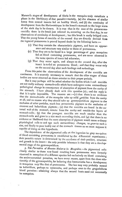
Manson's stages of development of filaria in the mosquito truly constitute a
phase in the life-history of that parasite-namely, (a) the absence of similar
forms from control insects fed on healthy blood; and (b) the continuity of
development from the filaria-embryos in the insect's stomach to the large worm
of the sixth day in its tissues. It is true that in the case of the proteosoma-
coccidia there is the break just referred to, occurring on the first day, in our
observations of continuity of development; but this break is easily bridged over.
That the young forms of coccidia of the second day are directly derived from
the ingested proteosoma is proved beyond doubt by the following facts:-
(1) That they contain the characteristic pigment, and have an appear-
ance and structure very similar to those of proteosoma.
(2) That they are to be found in a large percentage of grey mosquitos
fed on blood containing proteosoma, and are never observed in
the same species of insect fed otherwise.
(3) That they occur again, and always on the second day, after the
insect is re-fed on proteosoma blood; and that they occur only
on the second day after such feedings and re-feedings.
From this point the observations of the development of the coccidia are
continuous. It is scarcely necessary to remark that the older stages of these
bodies are never observed on dates anterior to their proper periods.
But it may perhaps still be asked whether the bodies which I call coccidia
are not really ordinary stomach-cells which have undergone some extraordinary
pathological change in consequence of absorption of pigment from the cavity of
the stomach. I have already dealt with this question (2); and the reply is
that it is quite impossible. The reasons are:-(1) that there is no evidence
that the stomach-cells of the mosquito take up solid particles from the cavity
at all, and no reason why they should take up gymnosporidium pigment to the
exclusion of other particles, much less proteosoma pigment to the exclusion of
crescent and halteridium pigment; (2) that the coccidia are found in the ex-
ternal wall of the stomach, remote from the cavity and outside the layers of
stomach-cells; (3) that the youngest coccidia are much smaller than the
stomach-cells and grow to a size much exceeding theirs, and (4) that there is no
evidence or likelihood that the mere absorption of pigment would cause ordinary
physiological cells to und rgo such extraordinary changes, to grow to such a
size, and finally to pass bodily out of the stomach tissues as we must suppose it
capable of doing on this hypothesis.
The dependence of the pigmented cells on the ingestion by grey mosquitos
of blood containing proteosoma is established by the differential experiments;
their parasitic nature is determined by the accidents of their position, structure
and growth in the insect: the only possible inference is that they are a develop-
mental stage of the gymnosporidium.
3. The Parasites of Human Malaria in Mosquitos.-As pigmented cells
closely similar to those now found resulting from proteosoma were formerly
observed in mosquitos of a different species fed exclusively on blood containing
the stivo-autumnal parasites, we have every reason, apart from the close rela-
tionship of the gymnosporidia, for believing that hmamba has a development
in mosquitos very like that of proteosoma. The law may very probably extend
to halteridium, perhaps to cytamba, and perhaps even to the gregariniform
blood parasites: admitting always that the second hosts need not necessarily
be mosquitos.
Set display mode to: Large image | Zoom image | Transcription
Images and transcriptions on this page, including medium image downloads, may be used under the Creative Commons Attribution 4.0 International Licence unless otherwise stated. ![]()
| India Papers > Medicine - Disease > Report on the cultivation of protesoma, Labbé, in grey mosquitoes > (26) Page 18 |
|---|
| Permanent URL | https://digital.nls.uk/74580868 |
|---|


