Encyclopaedia Britannica > Volume 3, Anatomy-Astronomy
(73) Page 65
Download files
Complete book:
Individual page:
Thumbnail gallery: Grid view | List view
ANATOMY.
65
Special The blood contained in the right chambers is modena-
Anatomy. coloured or venous; that of the left chambers is scarlet-
red or arterial. The former is derived from the vence
Course of which open into the right auricle; the latter from
throu h the Pulmonary veins> which open into the left. The di-
the°car(liacrection in which the blood flows on both sides is from the
diambs. venous apertures into the auricles, thence to the ventricles,
and thence to the respective arteries. The venous blood,
on reaching the auricle, distends it, and impels its muscles
to contraction ; and the cavity thus diminished expels the
blood in the only direction in which it can proceed,—by
the aurico-ventricular aperture into the ventricle. This
chamber being distended, its muscular walls all round,
especially at the base, contract and diminish its cavity,
when the blood, extruded, quits the ventricle in the only
direction in which it can, viz. by the aperture of the pul¬
monary artery. The blood from the pulmonary veins fol¬
lows the same course in the chambers of the left side.
The blood of the ventricles is prevented from returning
into the auricles partly by the tricuspid and mitral valves,
but chiefly by the annular contraction of the auriculo-ven-
tricular apertures, which are drawn from the margins to¬
wards the septum; while the latter is shortened, and the
apex is made to approach the base.
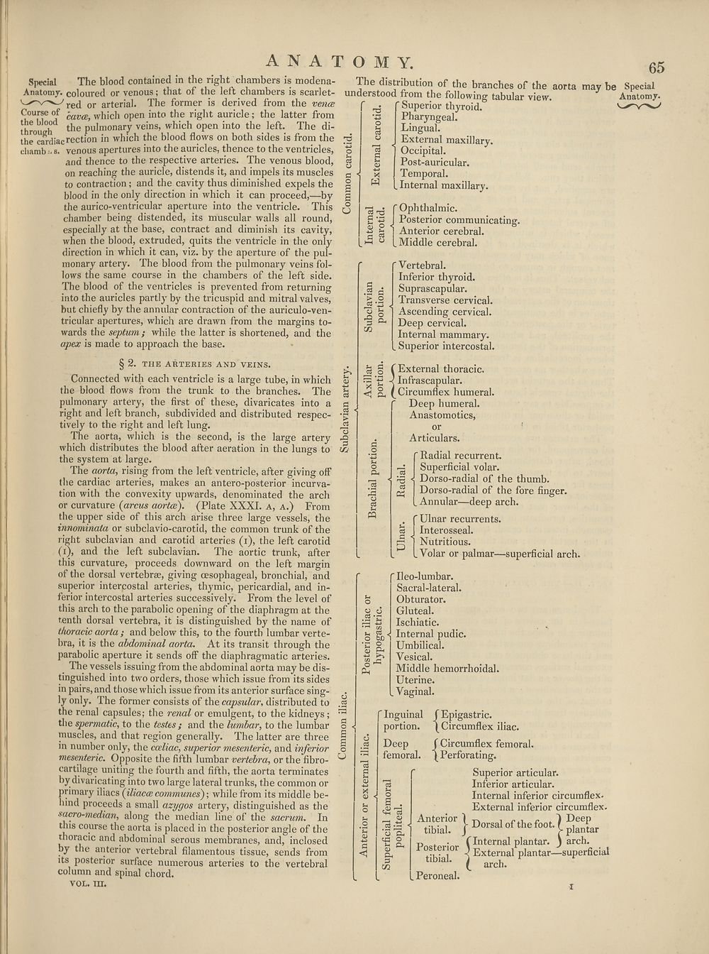
ihe distribution of the branches of the aorta may
understood fiom the following tabular view.
S
o
r
C3
□
u
tu
•*->
X
W
Superior thyroid.
Pharyngeal.
Lingual.
External maxillary.
Occipital.
Post-auricular.
Temporal.
.Internal maxillary.
15^ f Ophthalmic.
£ '-g I Posterior communicating,
•g £ 1 Anterior cerebral.
° [Middle cerebral.
'Vertebral.
Inferior thyroid.
Suprascapular.
Transverse cervical.
Ascending cervical.
Deep cervical.
Internal mammary.
. Superior intercostal.
§ 2. THE ARTERIES AND VEINS.
Connected with each ventricle is a large tube, in which
the blood flows from the trunk to the branches. The
pulmonary artery, the first of these, divaricates into a
right and left branch, subdivided and distributed respec¬
tively to the right and left lung.
The aorta, which is the second, is the large artery
which distributes the blood after aeration in the lungs to
the system at large.
The aorta, rising from the left ventricle, after giving off
the cardiac arteries, makes an antero-posterior incurva¬
tion with the convexity upwards, denominated the arch
or curvature (arcus aortce). (Plate XXXI. a, a.) From
the upper side of this arch arise three large vessels, the
innominata or subclavio-carotid, the common trunk of the
right subclavian and carotid arteries (i), the left carotid
(i), and the left subclavian. The aortic trunk, after
this curvature, proceeds downward on the left margin
of the dorsal vertebrae, giving oesophageal, bronchial, and
superior intercostal arteries, thymic, pericardial, and in¬
ferior intercostal arteries successively. From the level of
this arch to the parabolic opening of the diaphragm at the
tenth dorsal vertebra, it is distinguished by the name of
thoracic aorta; and below this, to the fourth lumbar verte¬
bra, it is the abdominal aorta. At its transit through the
parabolic aperture it sends off the diaphragmatic arteries.
The vessels issuing from the abdominal aorta may be dis¬
tinguished into two orders, those which issue from its sides
in pairs, and those which issue from its anterior surface sing¬
ly only. The former consists of the capsular, distributed to
the renal capsules; the renal or emulgent, to the kidneys;
the spermatic, to the testes; and the lumbar, to the lumbar
muscles, and that region generally. The latter are three
in number only, the cceliac, superior mesenteric, and inferior
mesenteric. Opposite the fifth lumbar vertebra, or the fibro-
cartilage uniting the fourth and fifth, the aorta terminates
by divaricating into two large lateral trunks, the common or
primary iliacs (iliacce communes'); while from its middle be¬
hind proceeds a small azygos artery, distinguished as the
sacro-median, along the median line of the sacrum. In
this course the aorta is placed in the posterior angle of the
thoracic and abdominal serous membranes, and, inclosed
by the anterior vertebral filamentous tissue, sends from
its posterior surface numerous arteries to the vertebral
column and spinal chord.
vol. m.
-o
3
irj
< o.
2
CQ
i bJD 1
O
1 £
^5
Pi
External thoracic.
Infrascapular.
Circumflex humeral.
Deep humeral.
Anastomotics,
or
Articulars.
Radial i*ecurrent.
Superficial volar.
Dorso-radial of the thumb.
Dorso-radial of the fore finger.
Annular—deep arch.
. f Ulnar recurrents.
c3 1 Interosseal,
p j Nutritious.
L Volar or palmar—superficial arch.
Ileo-lumbar.
Sacral-lateral.
Obturator.
Gluteal.
Ischiatic.
Internal pudic.
Umbilical.
Vesical.
Middle hemorrhoidal.
Uterine.
Vaginal.
(Inguinal
portion.
f Epigastric.
(Circumflex iliac.
Deep f Circumflex femoral,
femoral. (Perforating.
Superior articular.
Inferior articular.
Internal inferior circumflex.
External inferior circumflex.
Anterior 1 ^ rr Deep
tibial. / Dorsal of the foot-1 plantar
T1 , . (Internal plantar. ) arch.
tibhal°r i External plantar—superficial
. Peroneal.
a;
Oh
3
c/i
arch.
i
65
Special The blood contained in the right chambers is modena-
Anatomy. coloured or venous; that of the left chambers is scarlet-
red or arterial. The former is derived from the vence
Course of which open into the right auricle; the latter from
throu h the Pulmonary veins> which open into the left. The di-
the°car(liacrection in which the blood flows on both sides is from the
diambs. venous apertures into the auricles, thence to the ventricles,
and thence to the respective arteries. The venous blood,
on reaching the auricle, distends it, and impels its muscles
to contraction ; and the cavity thus diminished expels the
blood in the only direction in which it can proceed,—by
the aurico-ventricular aperture into the ventricle. This
chamber being distended, its muscular walls all round,
especially at the base, contract and diminish its cavity,
when the blood, extruded, quits the ventricle in the only
direction in which it can, viz. by the aperture of the pul¬
monary artery. The blood from the pulmonary veins fol¬
lows the same course in the chambers of the left side.
The blood of the ventricles is prevented from returning
into the auricles partly by the tricuspid and mitral valves,
but chiefly by the annular contraction of the auriculo-ven-
tricular apertures, which are drawn from the margins to¬
wards the septum; while the latter is shortened, and the
apex is made to approach the base.
ihe distribution of the branches of the aorta may
understood fiom the following tabular view.
S
o
r
C3
□
u
tu
•*->
X
W
Superior thyroid.
Pharyngeal.
Lingual.
External maxillary.
Occipital.
Post-auricular.
Temporal.
.Internal maxillary.
15^ f Ophthalmic.
£ '-g I Posterior communicating,
•g £ 1 Anterior cerebral.
° [Middle cerebral.
'Vertebral.
Inferior thyroid.
Suprascapular.
Transverse cervical.
Ascending cervical.
Deep cervical.
Internal mammary.
. Superior intercostal.
§ 2. THE ARTERIES AND VEINS.
Connected with each ventricle is a large tube, in which
the blood flows from the trunk to the branches. The
pulmonary artery, the first of these, divaricates into a
right and left branch, subdivided and distributed respec¬
tively to the right and left lung.
The aorta, which is the second, is the large artery
which distributes the blood after aeration in the lungs to
the system at large.
The aorta, rising from the left ventricle, after giving off
the cardiac arteries, makes an antero-posterior incurva¬
tion with the convexity upwards, denominated the arch
or curvature (arcus aortce). (Plate XXXI. a, a.) From
the upper side of this arch arise three large vessels, the
innominata or subclavio-carotid, the common trunk of the
right subclavian and carotid arteries (i), the left carotid
(i), and the left subclavian. The aortic trunk, after
this curvature, proceeds downward on the left margin
of the dorsal vertebrae, giving oesophageal, bronchial, and
superior intercostal arteries, thymic, pericardial, and in¬
ferior intercostal arteries successively. From the level of
this arch to the parabolic opening of the diaphragm at the
tenth dorsal vertebra, it is distinguished by the name of
thoracic aorta; and below this, to the fourth lumbar verte¬
bra, it is the abdominal aorta. At its transit through the
parabolic aperture it sends off the diaphragmatic arteries.
The vessels issuing from the abdominal aorta may be dis¬
tinguished into two orders, those which issue from its sides
in pairs, and those which issue from its anterior surface sing¬
ly only. The former consists of the capsular, distributed to
the renal capsules; the renal or emulgent, to the kidneys;
the spermatic, to the testes; and the lumbar, to the lumbar
muscles, and that region generally. The latter are three
in number only, the cceliac, superior mesenteric, and inferior
mesenteric. Opposite the fifth lumbar vertebra, or the fibro-
cartilage uniting the fourth and fifth, the aorta terminates
by divaricating into two large lateral trunks, the common or
primary iliacs (iliacce communes'); while from its middle be¬
hind proceeds a small azygos artery, distinguished as the
sacro-median, along the median line of the sacrum. In
this course the aorta is placed in the posterior angle of the
thoracic and abdominal serous membranes, and, inclosed
by the anterior vertebral filamentous tissue, sends from
its posterior surface numerous arteries to the vertebral
column and spinal chord.
vol. m.
-o
3
irj
< o.
2
CQ
i bJD 1
O
1 £
^5
Pi
External thoracic.
Infrascapular.
Circumflex humeral.
Deep humeral.
Anastomotics,
or
Articulars.
Radial i*ecurrent.
Superficial volar.
Dorso-radial of the thumb.
Dorso-radial of the fore finger.
Annular—deep arch.
. f Ulnar recurrents.
c3 1 Interosseal,
p j Nutritious.
L Volar or palmar—superficial arch.
Ileo-lumbar.
Sacral-lateral.
Obturator.
Gluteal.
Ischiatic.
Internal pudic.
Umbilical.
Vesical.
Middle hemorrhoidal.
Uterine.
Vaginal.
(Inguinal
portion.
f Epigastric.
(Circumflex iliac.
Deep f Circumflex femoral,
femoral. (Perforating.
Superior articular.
Inferior articular.
Internal inferior circumflex.
External inferior circumflex.
Anterior 1 ^ rr Deep
tibial. / Dorsal of the foot-1 plantar
T1 , . (Internal plantar. ) arch.
tibhal°r i External plantar—superficial
. Peroneal.
a;
Oh
3
c/i
arch.
i
Set display mode to:
![]() Universal Viewer |
Universal Viewer | ![]() Mirador |
Large image | Transcription
Mirador |
Large image | Transcription
Images and transcriptions on this page, including medium image downloads, may be used under the Creative Commons Attribution 4.0 International Licence unless otherwise stated. ![]()
| Encyclopaedia Britannica > Encyclopaedia Britannica > Volume 3, Anatomy-Astronomy > (73) Page 65 |
|---|
| Permanent URL | https://digital.nls.uk/193758297 |
|---|
| Attribution and copyright: |
|
|---|---|
| Shelfmark | EB.16 |
|---|---|
| Description | Ten editions of 'Encyclopaedia Britannica', issued from 1768-1903, in 231 volumes. Originally issued in 100 weekly parts (3 volumes) between 1768 and 1771 by publishers: Colin Macfarquhar and Andrew Bell (Edinburgh); editor: William Smellie: engraver: Andrew Bell. Expanded editions in the 19th century featured more volumes and contributions from leading experts in their fields. Managed and published in Edinburgh up to the 9th edition (25 volumes, from 1875-1889); the 10th edition (1902-1903) re-issued the 9th edition, with 11 supplementary volumes. |
|---|---|
| Additional NLS resources: |
|