Encyclopaedia Britannica > Volume 3, Athens-BOI
(736) Page 724
Download files
Complete book:
Individual page:
Thumbnail gallery: Grid view | List view
724
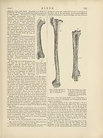
rudiment. Hence the Ostrich has only two toes (which
answer to the third and fourth of the pentadactyle foot),
with four phalanges in the inner and five in the outer,
though the inner toe is far the longer and the stronger.
In most four-toed Birds the hallux is turned more or less
completely backwards, and the other three digits forwards.
But in many Aetomorphce (especially the Owls) the outer
toe can be turned outwards, or even backwards, at will.
And in the Parrots, Toucans, Cuckoos, Woodpeckers, and
other so-called “ Scansorial ” Birds, the outer toe is per¬
manently reversed. Under these circumstances the distal
end of the outer metatarsal may be divided into two dis¬
tinct articular surfaces. In the Trogons there are two toes
in front and two behind, as in the Parrots; but it is the
second toe which is turned backwards. Lastly, in the
Swifts, the Dysporomorphce, and the Spheniscomorphce, the
hallux is directed more or less forwards, so that all four
toes are turned to the front.
As a general rule, the osseous tissue of Birds is remark¬
ably dense and hard. Before hatching, the bones are solid
and filled with vascular medulla; but after birth, more or
fewer of the bones are always excavated by prolongation
of cavities containing air, which lie in their neighbourhood.
Such air cavities are always found in the skull, in connec¬
tion with the nasal and auditory passages, and they may
extend through all parts of the skull, with the exception
of the jugal arch, which, however, is pneumatic in the
Toucan and Hornbill. In many birds, Apteryx, Penguin,
Divers (and Gulls, according to Professor Huxley; but this
is a mistake, their spinal column far into the sacrum is
pneumatic; Larus canus shows this well), and the smaller
Song-birds, no other bones than those of the skull are
pneumatic; but in most birds the air-sacs of the lungs
send prolongations into the bones of the rest of the trunk-
skeleton, seldom into the caudal vertebrae, as in Balceniceps,
the Adjutant, Hornbill, &c. In the Hornbills the whole
skeleton is pneumatic; in a large number of birds the
humerus alone of the limb-bones contains air; in the
diurnal Raptores, the femur also. It is proper to remark
that the amount of pneumaticity of bones by no means
follows the development of the powers of flight. In the
Ostrich, for example, the bones are far more extensively
pneumatic than in the Gull.
In some cases, prolongations of the air-sacs extend
beneath the integument.
The Muscles.
In the space allotted to the writer, there is merely room
for justice to be done to one category of organs ; and as the
skeleton, and especially the skull, is of most direct import¬
ance to the zoologist and palaeontologist, and as its form
determines, as it were, all other organs, they being correlated
with it and answering to it, it seemed to be that on which
election should fall for the fuller treatment. An impartial
description of all the systems of organs would have resulted
in the merest outline for each. For the muscles, Professor
Huxley’s abstract must serve.1
The cutaneous muscles of Birds are well developed, and
form broad expansions in various parts of the body. Special
1 Anat. Vert. Anim., p. 300. For an almost exhaustive biblio¬
graphy of writings on the muscular system of birds, see M. Edmond
Alix’s Essai sur l'Appareil locomoteur des Oiseaux, pp. 367-373.
This list begins with Aldrovandus, 1581, and ends with Goverod, 1873,
1874. We miss, however, Macgillivray’s excellent description, with
figures, of the muscles of flight, Brit. Birds, vol. i. plate 3, pp. 35-46;
and another by Professor Rolleston, “ On Muscles connected with the
Shoulder-joint,” Trans. Linn. Soc., vol. xxvi. pp. 610-629. See also
Owen “ On the Apteryx” Trans. Zool. Soc., vol. vii. p. 381, pi. 46.
But the most important work for reference is that of M. Alix himself
(op. cit., pp. 373-471, plates 1-3, “Appareil actif de la Locomotion ”).
[anatomy.
bundles of muscular fibres pass to the great quill feathers
of the tail and wings, and others to the patagium, a fold of
integument which stands between the trunk and brachium
behind and between the brachium and ante-brachium in
front. In correspondence with the slight mobility of the
dorsal vertebrae, the episkeletal and hyposkeletal muscles
of the spine attain a considerable development only in the
neck and in the tail. Owing to the great size of the
sternum, the abdominal muscles are usually small, and the
internal oblique may be absent. A diaphragm, consisting
of bundles of muscular fibre,2 which pass from the ribs to
the aponeurosis, covering the ventral face of the lungs, is
developed in all Birds, but attains to the greatest degree of
completeness in the Ratitoe, and especially in Apteryx.
The muscles of the limbs are remarkably modified by the
excessive development of some of those found in other
Vertebrata, and the suppression of others.
Thus in all birds possessing the power of flight, the pec-
toralis major, the chief agent of the downward stroke of
the wing, is very large and thick, taking its origin from the
whole length, and a great part of the depth, of the keel of
the sternum. The elevation of the wing is chiefly effected
by the pectoralis secundus (levator humeri; or p. medius,
Macg., plate 3, figs. 4, 5), which arises beneath (within and
over, in the standing bird) the foregoing muscle, and passes
over the inner side of the scapulo-coracoid articulation as
over a pulley, to reach the humerus. The muscles of the
fore-arm and digits are reduced, in accordance with the
peculiar modification of the skeleton of these parts. In
the hind limb of most birds there is a singular extensor
muscle, which arises from the pubis, and ends in a tendon
which passes to the outside of the knee-joint and terminates
in the leg by uniting with the yfecor digitorum perf oratus.
The result of this arrangement is that the toes are flexed
whenever the leg is bent upon the thigh, and consequently
the roosting bird is held fast upon his perch by the weight
of his own body.3
The Brain.
In Birds, as in Reptiles, the cerebro-spinal axis is angu-
lated at the junction of the spinal cord with the medulla
oblongata, the latter being bent down towards the ventral
side of the body. The region on which the nerves of
the anterior and posterior extremities originate is enlarged
in Birds. In the lumbar enlargement the posterior
columns of the cord diverge and give rise to the sinus
rhomboidalis, which is a sort of repetition of the fourth
ventricle, the dilated central canal of the spinal cord being
covered merely by a thin membrane, consisting chiefly of
the ependyma and arachnoid. The brain fills the cavity
of the skull, and presents a well-developed cerebellum; a
mesencephalon, divided above into two optic lobes; and
relatively large prosencephalic hemispheres, which attain a
considerable size but never conceal the optic lobes. The
transverse fissures of the cerebellum are distinct, and the
lateral appendages of the cerebellum, or jloccnli, become
well defined, and are wedged, as in many of the lower
Mammalia, in cavities of the side walls of the skull, arched
over by the anterior vertical semicircular canal.
There is no pons Varolii, in the sense of transverse fibres
connecting the two halves of the cerebellum, visible upon
the ventral surface of the mesencephalon. The optic lobes
contain ventricles; these are thrown down to the sides of
the base of the brain, and are connected over the aquaeductus
Sylvii by a broad commissural band. Each prosencephalic
lobe contains a lateral ventricle (continuous through the
2 See Macgillivray, Brit. Birds, vol. ii. plate 11, fig. 1, v.v.v.
3 See J. Alph. Borelli, De Motu Animalium, Romas, 1680-1682,
Lugd. Bat. 1865 ; and Bibliotheca Anatomica, Geneva, 1685, plate 82,
figs. 4-7.
BIRDS
Set display mode to:
![]() Universal Viewer |
Universal Viewer | ![]() Mirador |
Large image | Transcription
Mirador |
Large image | Transcription
Images and transcriptions on this page, including medium image downloads, may be used under the Creative Commons Attribution 4.0 International Licence unless otherwise stated. ![]()
| Encyclopaedia Britannica > Encyclopaedia Britannica > Volume 3, Athens-BOI > (736) Page 724 |
|---|
| Permanent URL | https://digital.nls.uk/193659869 |
|---|
| Attribution and copyright: |
|
|---|---|
| Shelfmark | EB.17 |
|---|---|
| Description | Ten editions of 'Encyclopaedia Britannica', issued from 1768-1903, in 231 volumes. Originally issued in 100 weekly parts (3 volumes) between 1768 and 1771 by publishers: Colin Macfarquhar and Andrew Bell (Edinburgh); editor: William Smellie: engraver: Andrew Bell. Expanded editions in the 19th century featured more volumes and contributions from leading experts in their fields. Managed and published in Edinburgh up to the 9th edition (25 volumes, from 1875-1889); the 10th edition (1902-1903) re-issued the 9th edition, with 11 supplementary volumes. |
|---|---|
| Additional NLS resources: |
|