Encyclopaedia Britannica > Volume 3, Athens-BOI
(734) Page 722
Download files
Complete book:
Individual page:
Thumbnail gallery: Grid view | List view
[anatomy.
722
BIRDS
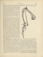
a less degree, in the Great Auk, the humerus becomes
ilattened from side to side, the proximal end is singularly
modified, and at the narrow distal end the articular sur¬
face for the radius lies completely in front of, and rather
above, that for the ulna.
The ulna, which often presents a series of tubercles,
indicating the attachment of the secondary quill feathers,
is usually a stronger and a longer bone than the radius.
There are only two carpal bones, one radial and one ulnar.
There is one exception to this, namely, in the Screamer
(Chauna chavaria), which has three carpals on the left side,
the lower arcuate bone having two representatives.
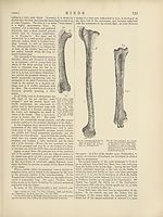
In the Apterygidce and in the Casuariidce there is but
one complete digit in the manus. It appears to answer to
the second of the pentadactyle limb, and is provided with
a claw. In the Strutkionidce and liheidce, and in all the
Carinatce, there are three digits in the manus, which
answer to the pollex and the second and third digits of
the pentadactyle fore-limb; and the metacarpal bones of
these digits are ankylosed together. As a rule the meta¬
carpal of the pollex is much shorter than the other two;
that of the second digit is strong and straight; that of the
third is more slender and bowed, so as to leave an inter¬
space between itself and the second, which is often filled
up by bony matter. The pollex has two phalanges, and
the second of them is, in many birds—Rhea, the Screamer,
<fec.—pointed, curved, and ensheathed in a horny claw.
The second digit has two and sometimes three phalanges,
as in the Swan ; and the terminal phalanx is similarly pro¬
vided with a claw in sundry birds, e.g., the Swan and Rhea.
In the Ostrich both the pollex and the second digit are
unguiculate. The third digit possesses one phalanx, besides
it's ankylosed metacarpal, and is always devoid of a claw.
It is a singular circumstance that the relative proportions
of the humerus and the manus should present the most
marked contrast in two groups of birds which are alike
remarkable for their powers of flight. These are the
Swifts and Humming-birds, in which the humerus is short
and the manus long, and the Albatrosses, in which the
humerus is long and the manus relatively short.
In the Penguins the pollex has two free phalanges, and
its metacarpal bone (which is distinct in the young birds)
ankyloses with that of the second digit. The third
metacarpal is slender and straight. The bones of the
manus are singularly elongated and flattened.
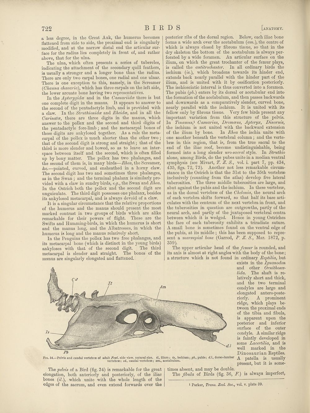
posterior ribs of the dorsal region. Below, each iliac bone
forms a wide arch over the acetabulum {am.), the centre of
which is always closed by fibrous tissue, so that in the
dry skeleton the bottom of the acetabulum is always per¬
forated by a wide foramen. An articular surface on the
ilium, on which the great trochanter of the femur plays,
is called the antitrochanter. In all ordinary birds the
ischium (fs.), which broadens towards its hinder end,
extends back nearly parallel with the hinder part of the
ilium, and is united with it by ossification posteriorly.
The ischiosciatic interval is thus converted into a foramen.
The pubis (gpb.) enters by its dorsal or acetabular end into
the formation of the acetabulum, and then passes backwards
and downwards as a comparatively slender, curved bone,
nearly parallel with the ischium. It is united with its
fellow only by fibrous tissue. Very few birds present any
important variation from this structure of the pelvis.
In Tmamus,1 Casuarius, Dromoeus, Apteryx, Dinornis,
the ischium is not united with the backward extension
of the ilium by bone. In Rhea the ischia unite with
one another beneath the vertebral column; and the verte¬
brae in this region, that is, from the true sacral to the
end of the iliac roof, become undistir guishable, being
formed into a long slender uro-sacral style. In Struthio
alone, among Birds, do the pubes unite in a median ventral
symphysis (see Mivart, T. Z. S., vol. i. part 7, pp. 434,
435, figs. 72, 73). Another not less remarkable circum¬
stance in the Ostrich is that the 31st to the 35th vertebrae
inclusively (counting from the atlas) develop five lateral
tuberosities. The three middle tuberosities are large, and
abut against the pubis and the ischium. In these vertebrae,
as in the dorsal vertebrae of the Chelonia, the neural arch
of each vertebra shifts forward, so that half its base arti¬
culates with the centrum of the next vertebra in front, and
the tuberosities in question are outgrowths, partly of the
neural arch, and partly of the juxtaposed vertebral centra
between which it is wedged. Hence in young Ostriches
the face of each tuberosity exhibits a triradiate suture.
A small bone is sometimes found on the ventral edge of
the pubis, at its middle ; this has been supposed to repre¬
sent a marsupial bone (Garrod, P. Z. S., Mar. 1872, p.
359).
The upper articular head of the femur is rounded, and
its axis is almost at right angles with the body of the bone;
a structure which is not found in ordinary Reptilia, but
exists in the Iguanodon
and other Ornithosce-
Uda. The shaft is re¬
latively short and thick,
and the two terminal
condyles are large and
elongated antero-poste-
riorly. A prominent
ridge, which plays be¬
tween the proximal ends
of the tibia and fibula,
is apparent upon the
posterior and inferior
surface of the outer
condyle. A similar ridge
is faintly developed in
^ some Lacertilia, and is
well marked in the
Dinosaurian Reptiles.
t7., ilium; ts., ischium; p&., pubis; cU., dorse-lumbar ^ natella is usually
am., acetabulum. r . J
present, but it is some-
Pi
Fig. 34.—Pelvis and caudal vertebrse of adult Fowl, side view, natural size.
vertebrae; cd., caudal vertebrae;
The pelvis of a Bird (fig. 34) is remarkable for the great
elongation, both anteriorly and posteriorly, of the iliac
bones {il.), which unite with the whole length of the
edges of the sacrum, and even extend forwards over the
times absent, and may be double.
The fibula of Birds (fig. 36, F.) is always imperfect,
i Parker, Trans. Zool. Soc., vol. v. plate 39.
722
BIRDS
a less degree, in the Great Auk, the humerus becomes
ilattened from side to side, the proximal end is singularly
modified, and at the narrow distal end the articular sur¬
face for the radius lies completely in front of, and rather
above, that for the ulna.
The ulna, which often presents a series of tubercles,
indicating the attachment of the secondary quill feathers,
is usually a stronger and a longer bone than the radius.
There are only two carpal bones, one radial and one ulnar.
There is one exception to this, namely, in the Screamer
(Chauna chavaria), which has three carpals on the left side,
the lower arcuate bone having two representatives.
In the Apterygidce and in the Casuariidce there is but
one complete digit in the manus. It appears to answer to
the second of the pentadactyle limb, and is provided with
a claw. In the Strutkionidce and liheidce, and in all the
Carinatce, there are three digits in the manus, which
answer to the pollex and the second and third digits of
the pentadactyle fore-limb; and the metacarpal bones of
these digits are ankylosed together. As a rule the meta¬
carpal of the pollex is much shorter than the other two;
that of the second digit is strong and straight; that of the
third is more slender and bowed, so as to leave an inter¬
space between itself and the second, which is often filled
up by bony matter. The pollex has two phalanges, and
the second of them is, in many birds—Rhea, the Screamer,
<fec.—pointed, curved, and ensheathed in a horny claw.
The second digit has two and sometimes three phalanges,
as in the Swan ; and the terminal phalanx is similarly pro¬
vided with a claw in sundry birds, e.g., the Swan and Rhea.
In the Ostrich both the pollex and the second digit are
unguiculate. The third digit possesses one phalanx, besides
it's ankylosed metacarpal, and is always devoid of a claw.
It is a singular circumstance that the relative proportions
of the humerus and the manus should present the most
marked contrast in two groups of birds which are alike
remarkable for their powers of flight. These are the
Swifts and Humming-birds, in which the humerus is short
and the manus long, and the Albatrosses, in which the
humerus is long and the manus relatively short.
In the Penguins the pollex has two free phalanges, and
its metacarpal bone (which is distinct in the young birds)
ankyloses with that of the second digit. The third
metacarpal is slender and straight. The bones of the
manus are singularly elongated and flattened.
posterior ribs of the dorsal region. Below, each iliac bone
forms a wide arch over the acetabulum {am.), the centre of
which is always closed by fibrous tissue, so that in the
dry skeleton the bottom of the acetabulum is always per¬
forated by a wide foramen. An articular surface on the
ilium, on which the great trochanter of the femur plays,
is called the antitrochanter. In all ordinary birds the
ischium (fs.), which broadens towards its hinder end,
extends back nearly parallel with the hinder part of the
ilium, and is united with it by ossification posteriorly.
The ischiosciatic interval is thus converted into a foramen.
The pubis (gpb.) enters by its dorsal or acetabular end into
the formation of the acetabulum, and then passes backwards
and downwards as a comparatively slender, curved bone,
nearly parallel with the ischium. It is united with its
fellow only by fibrous tissue. Very few birds present any
important variation from this structure of the pelvis.
In Tmamus,1 Casuarius, Dromoeus, Apteryx, Dinornis,
the ischium is not united with the backward extension
of the ilium by bone. In Rhea the ischia unite with
one another beneath the vertebral column; and the verte¬
brae in this region, that is, from the true sacral to the
end of the iliac roof, become undistir guishable, being
formed into a long slender uro-sacral style. In Struthio
alone, among Birds, do the pubes unite in a median ventral
symphysis (see Mivart, T. Z. S., vol. i. part 7, pp. 434,
435, figs. 72, 73). Another not less remarkable circum¬
stance in the Ostrich is that the 31st to the 35th vertebrae
inclusively (counting from the atlas) develop five lateral
tuberosities. The three middle tuberosities are large, and
abut against the pubis and the ischium. In these vertebrae,
as in the dorsal vertebrae of the Chelonia, the neural arch
of each vertebra shifts forward, so that half its base arti¬
culates with the centrum of the next vertebra in front, and
the tuberosities in question are outgrowths, partly of the
neural arch, and partly of the juxtaposed vertebral centra
between which it is wedged. Hence in young Ostriches
the face of each tuberosity exhibits a triradiate suture.
A small bone is sometimes found on the ventral edge of
the pubis, at its middle ; this has been supposed to repre¬
sent a marsupial bone (Garrod, P. Z. S., Mar. 1872, p.
359).
The upper articular head of the femur is rounded, and
its axis is almost at right angles with the body of the bone;
a structure which is not found in ordinary Reptilia, but
exists in the Iguanodon
and other Ornithosce-
Uda. The shaft is re¬
latively short and thick,
and the two terminal
condyles are large and
elongated antero-poste-
riorly. A prominent
ridge, which plays be¬
tween the proximal ends
of the tibia and fibula,
is apparent upon the
posterior and inferior
surface of the outer
condyle. A similar ridge
is faintly developed in
^ some Lacertilia, and is
well marked in the
Dinosaurian Reptiles.
t7., ilium; ts., ischium; p&., pubis; cU., dorse-lumbar ^ natella is usually
am., acetabulum. r . J
present, but it is some-
Pi
Fig. 34.—Pelvis and caudal vertebrse of adult Fowl, side view, natural size.
vertebrae; cd., caudal vertebrae;
The pelvis of a Bird (fig. 34) is remarkable for the great
elongation, both anteriorly and posteriorly, of the iliac
bones {il.), which unite with the whole length of the
edges of the sacrum, and even extend forwards over the
times absent, and may be double.
The fibula of Birds (fig. 36, F.) is always imperfect,
i Parker, Trans. Zool. Soc., vol. v. plate 39.
Set display mode to:
![]() Universal Viewer |
Universal Viewer | ![]() Mirador |
Large image | Transcription
Mirador |
Large image | Transcription
Images and transcriptions on this page, including medium image downloads, may be used under the Creative Commons Attribution 4.0 International Licence unless otherwise stated. ![]()
| Encyclopaedia Britannica > Encyclopaedia Britannica > Volume 3, Athens-BOI > (734) Page 722 |
|---|
| Permanent URL | https://digital.nls.uk/193659843 |
|---|
| Attribution and copyright: |
|
|---|---|
| Shelfmark | EB.17 |
|---|---|
| Description | Ten editions of 'Encyclopaedia Britannica', issued from 1768-1903, in 231 volumes. Originally issued in 100 weekly parts (3 volumes) between 1768 and 1771 by publishers: Colin Macfarquhar and Andrew Bell (Edinburgh); editor: William Smellie: engraver: Andrew Bell. Expanded editions in the 19th century featured more volumes and contributions from leading experts in their fields. Managed and published in Edinburgh up to the 9th edition (25 volumes, from 1875-1889); the 10th edition (1902-1903) re-issued the 9th edition, with 11 supplementary volumes. |
|---|---|
| Additional NLS resources: |
|